 Fig. 1
 Fig. 2
 Fig. 3
 Fig. 4
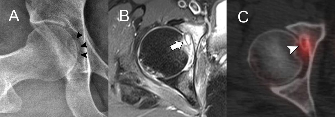
A 43-year-old woman presented with 2 months of localized pain in the right hip after a fall from standing height. The pain was aggravated with walking. On physical examination, there was no localized osseous tenderness except for a positive Patrick (FABER [Flexion, Abduction, and External Rotation]) test, and internal rotation was restricted to 15° compared with 30° on the contralateral side. Imaging included radiographs, magnetic resonance imaging (MRI), and single-photon emission computed tomography (SPECT/CT) (Fig. 1). Initial radiographs showed no evidence of fracture, but a subtle radiopaque lesion was seen at the acetabular fossa. MRI revealed a 1-cm mass-like lesion in the anterior part of the acetabular fossa, protruding into the right hip joint with adjacent bone marrow edema. SPECT/CT showed focal hot uptake at the same site. An arthroscopic excision was planned.
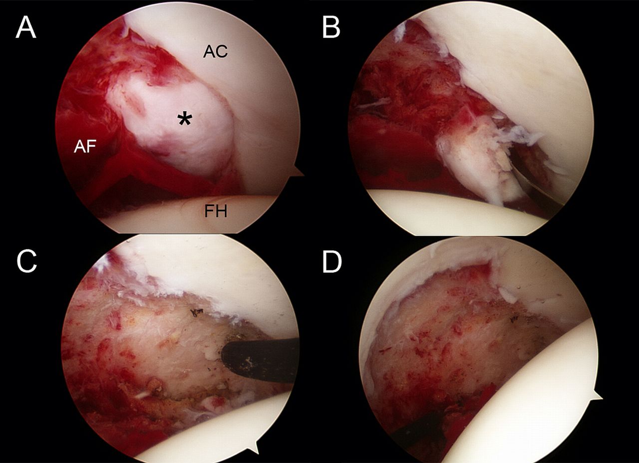
With the patient in the supine position, we used a standard 3-portal technique with a commercially available hip arthroscopy distraction unit (Smith & Nephew). With the limb in traction, we first examined the central compartment using a 70° scope. On initial exploration, a diffuse hematoma and synovitis were seen at the acetabular fossa. After hematoma evacuation, a whitish solitary cartilaginous lesion was identified along the anterior border of the acetabular notch (zone 6 according to the geographic zone method). For better visualization and instrument manipulation, an anterior capsulotomy was performed. After complete exposure of the borders of the mass with the use of a 30° scope, the mass was excised en bloc using an arthroscopic elevator and a pituitary rongeur, and it was sent for histopathologic examination. Radiofrequency (RF) thermal ablation of the margin was performed with use of a flexible RF probe (VULCAN; Smith & Nephew). After debridement and synovectomy of the fossa, complete excision of the lesion was confirmed with arthroscopic examination of the acetabular fossa using both the 70° and 30° scopes (Fig. 2). The labrum, articular cartilage of the femur, and the acetabulum were normal on arthroscopic examination.
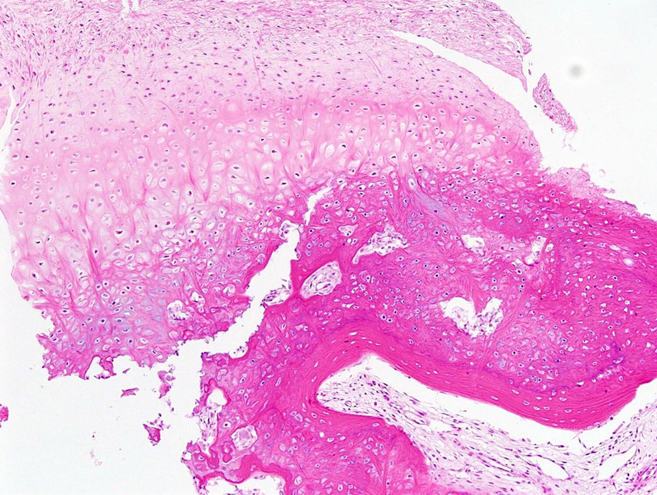
The histopathology of the excised lesion is shown in Figure 3.
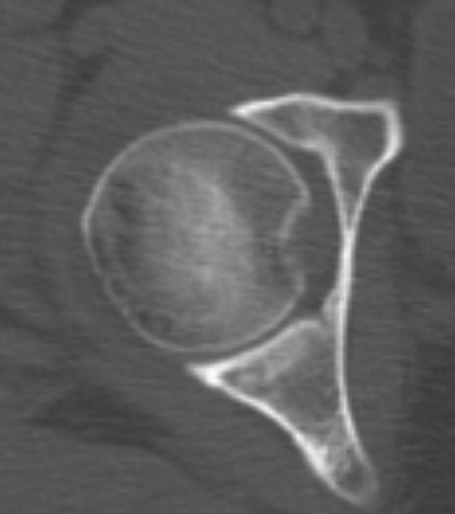
The patient was diagnosed with an osteochondroma. The patient was allowed weight-bearing with crutches as tolerated and started continuous passive motion exercises on postoperative day 1. She was discharged 2 days after surgery. At the first follow-up visit (6 weeks postoperatively), the patient was asymptomatic and there was normal range of motion of the right hip (internal rotation was 30° for both hips). CT performed 3 months postoperatively confirmed that there had been complete excision of the osteochondroma (Fig. 4). At the 1-year follow-up, the patient was asymptomatic, and there was no evidence of local recurrence on radiographic examination.
Proceed to Discussion >>Reference: Kim CH, Kekatpure AL, Kashikar A, Chang JS, Jeong MY, Yoon PW. Arthroscopic excision of a solitary acetabular osteochondroma in an adult: a case report. JBJS Case Connect, 2016 Dec 28;6(4):e101.
Periarticular osteochondromas around the hip are relatively rare, and most of them have been reported in association with hereditary multiple exostoses. They are commonly located in the peritrochanteric region but are rare in the acetabular fossa. Such a solitary lesion is often diagnosed with detailed imaging of the hip, including CT and MRI when the standard anteroposterior and lateral radiographs do not reveal substantial osseous abnormality.
Hip arthroscopy has various diagnostic and therapeutic indications in adults and is used mainly for the management of femoroacetabular impingement, postinjury removal of osteochondral fracture fragments, labral repair, and internal and external snapping hip syndrome. It has several advantages over open surgical dislocation in the management of intra-articular lesions, including being minimally invasive with minimal damage to the femoral head blood supply and allowing early rehabilitation. In the literature, we found 8 previously reported cases of acetabular osteochondroma in young patients that were treated mostly with open excision; there was only 1 case report describing arthroscopic treatment of acetabular osteochondroma in 2 children with hereditary multiple exostoses. Recently, a few cases of arthroscopic excision of osteochondroma of the femoral neck have been reported, but to our knowledge, this is the first case report of arthroscopic excision of an acetabular osteochondroma in an adult with no history of hereditary multiple exostoses. Surgeons should be aware that an osteochondroma can occur at the acetabular fossa as a solitary lesion, even in an adult, and hip arthroscopy can be a good option for the management of an intra-articular solitary lesion of the hip without risk of damage to the blood supply of the femoral head.
Reference: Kim CH, Kekatpure AL, Kashikar A, Chang JS, Jeong MY, Yoon PW. Arthroscopic excision of a solitary acetabular osteochondroma in an adult: a case report. JBJS Case Connect, 2016 Dec 28;6(4):e101.
What is the diagnosis?
Osteocartilaginous loose body
Osteochondritis dissecans
Bizarre parosteal osteochondromatous proliferation (Nora lesion)
Osteochondroma
Enchondroma