A Child with Unremarkable Gestation Born with Dysmorphic Features
March 20, 2019
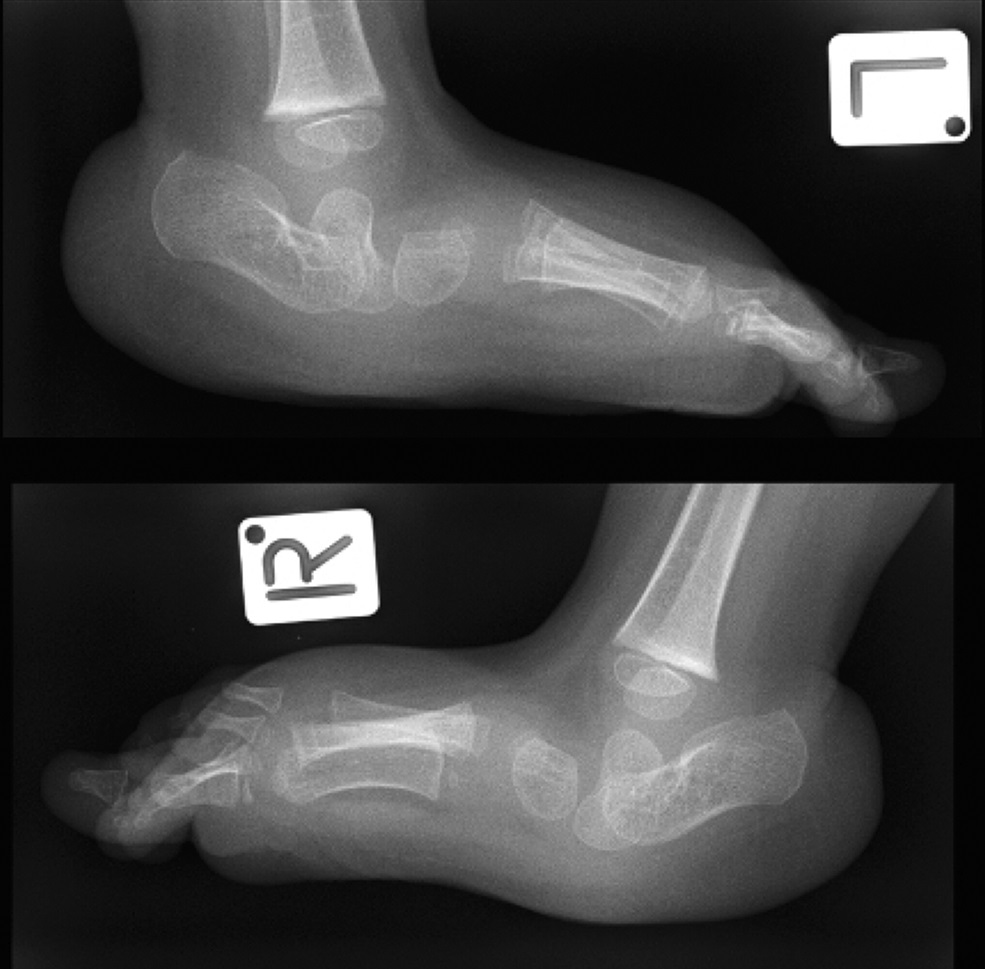
 Fig. 1
 Fig. 2
 Fig. 3
 Fig. 4
 Fig. 5
The child was a firstborn who had been delivered to healthy parents by cesarean section at 40 weeks after failure to progress. Clomiphene stimulation had been used because of maternal polycystic ovarian syndrome, but the pregnancy was otherwise unremarkable, and an ultrasound scan of the anatomy at 19 weeks’ gestation was normal.
Dysmorphic features were noted at birth, including contractures of the fingers with an overlapping thumb and a little finger that overlapped the ring and middle fingers. There were dorsiflexion contractures of the feet, and both hips were dislocated. Furthermore, it was noted that there was limited extension of the knees and elbows and that the heels were prominent. An atrial septal defect was detected on echocardiography. The ears had a truncated superior helix and an overfolded appearance. Based on these clinical features, a diagnosis of Beals contractural arachnodactyly was made at 14 weeks postpartum by a pediatric geneticist.
The finger contractures were managed with gentle stretching and manipulation and had a good outcome. Management of the dislocated hips involved an attempt to treat with a Pavlik harness, which was unsuccessful as seen on sequential ultrasound scans that demonstrated persistent dislocation; subsequent bilateral open reduction was required when the patient was 1 year of age. Radiographs of the feet when the child was 2 years of age are shown in Figure 1.
The clinical and radiographic features led to the diagnosis of congenital vertical talus (CVT).
At 2 years of age, the patient underwent correction of bilateral CVT in sequential operations. The talus was reduced in a single-stage operation that utilized 3 incisions: a lateral incision that was centered over the sinus tarsi, a medial incision that was centered over the head of the talus, and an incision medial to the Achilles tendon. Once the talus was reduced, a sling was created by splitting the tibialis anterior, passing it around the neck of the talus, and rejoining it to provide dynamic stability. Z-lengthening of the peroneal tendons, the extensor tendons of the toes, and the Achilles tendon also was undertaken via an extension of the lateral incision (Fig. 2).
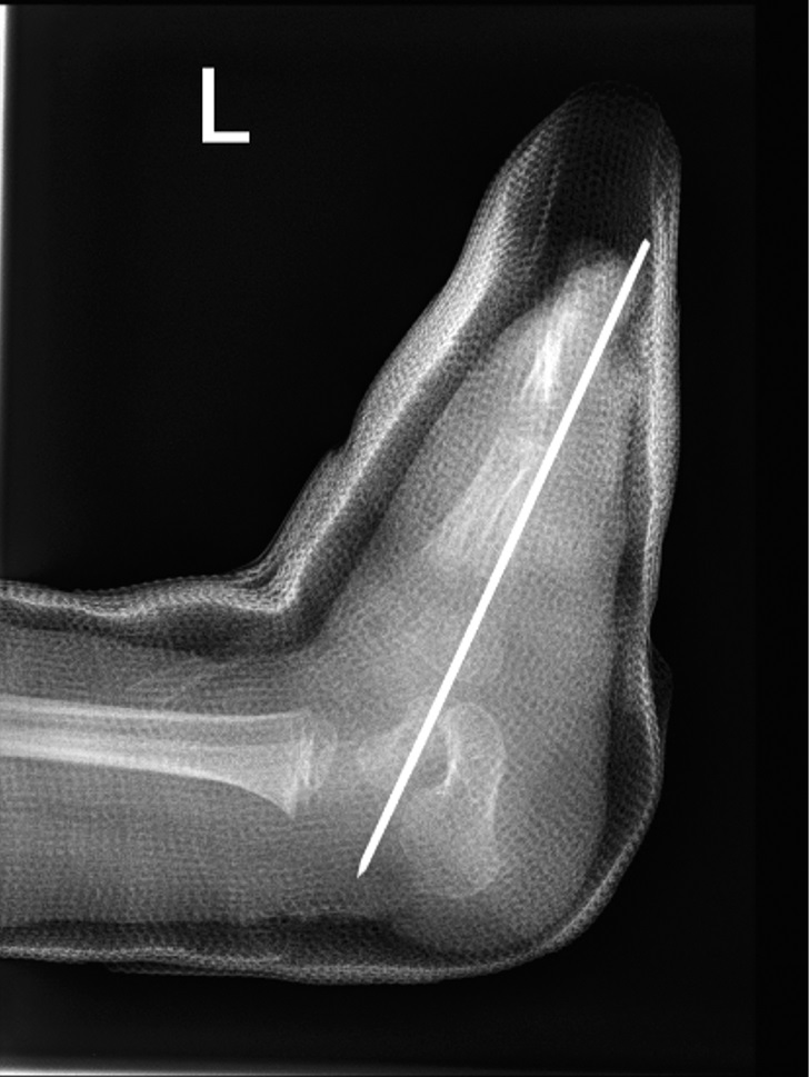
After a year, the malalignment of the left talus recurred (Fig. 3), and subsequent surgery was performed. During the revision operation, in addition to detachment of the tibialis anterior construct, extensive scarring was noted. After release, the navicular was mobilized onto the head of the talus, and a Kirschner wire was used to hold the position, as described by Dobbs et al. Furthermore, the tibialis anterior tendon was augmented with a tendon of the extensor digitorum longus. This was divided distally to maintain integrity of the muscle-bone unit, and it was transferred to the neck of the talus and held with a titanium suture anchor.
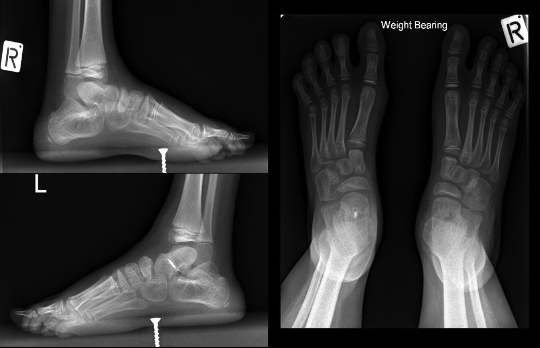
The most recent follow-up was when the patient was 7 years old. Although there was some slight residual planovalgus deformity, the child was walking independently with no notable gait disturbance, no substantial ankle and subtalar joint stiffness, and no functional limitation compared with peers. The final foot alignment is shown in Figures 4 and 5.
Proceed to Discussion >>Reference: Morris H, Navarre P. Bilateral congenital vertical talus in association with Beals contractural arachnodactyly: a case report. JBJS Case Connect. 2018 Oct-Dec;8(4):e97.
Beals syndrome, also known as congenital contractural arachnodactyly, is an autosomal-dominant arthrogryposis that is characterized by kyphoscoliosis, abnormality of the external aspect of the ears, dolichostenomelia, arachnodactyly, and multiple contractures. Although the joint contractures are congenital, they have a tendency to lessen with age and with growth of the child, but interventions often are needed.
Historically, Beals contractural arachnodactyly often was misdiagnosed as Marfan syndrome because of an overlap of characteristic features. While both syndromes are due to a defect in the fibrillin gene, each syndrome is caused by a defect at a differing genetic locus. Marfan syndrome is caused by a defect in the fibrillin-1 gene at 15q15-21, whereas Beals syndrome is caused by a defect in the fibrillin-2 gene at 5q23-31. Fibrillin-2 is a component of microfibrils, which are found in tissues that are associated with the elastin system. Fibrillin-2 appears with elastogenesis early in development, and it is predominantly located in elastic tissues (e.g., elastic cartilage and the tunica media of the aorta). In mice, it has been shown that an absence of the fibrillin-2 gene alters the constituents of tendons and causes an abnormality in bone growth, thus suggesting that fibrillin-2 influences the biochemical and morphological processes in connective tissue.
CVT has been described in a range of neuromuscular conditions, including myelodysplasia and arthrogryposis. The literature on CVT reveals conflicting pathoanatomy and recommended management of the abnormality. Nevertheless, a unifying feature among reported cases is substantial pathology that is located at the level of the subtalar joint. If untreated, the natural history is pain, abnormal callus formation, issues with shoe wear, and an abnormal gait. Treatment aims to restore normal relationships between the talus, the navicular, and the calcaneus, and to provide a plantigrade weight-bearing surface on the sole of the foot.
There are characteristic radiographic appearances with CVT. Standard views include an anteroposterior view and 3 lateral views: with the foot in maximal dorsiflexion, maximal plantar flexion, and neutral. Standing views can be made with older children; on a weight-bearing lateral view, the talus appears in a nearly vertical position, almost parallel to the tibia. Hamanishi defined criteria for the radiographic diagnosis of CVT. In maximum plantar flexion, a talar axis-first metatarsal base angle (TAMBA) of >30° is diagnostic of CVT. Ultrasonography may be used to diagnose CVT in infancy, and there are reports of the use of magnetic resonance imaging (MRI) for studying the pathoanatomy of the condition.
Traditionally, correction in patients who are <2 years old has been by surgical methods with a 1 or 2-stage extensive soft-tissue release. The first stage of the 2-stage approach included lengthening of the dorsolateral tendons, and the second stage involved lengthening of the Achilles and peroneal tendons. The 1-stage approach was a combination of these 2 stages. Complications include stiffness and degenerative osteoarthritis, which may require salvage procedures such as subtalar and triple arthrodeses. There have been reports of additional procedures that include excision of the navicular and arthrodesis of the subtalar joint.
In 2006, a new method referred to as the reverse Ponseti, or Dobbs, technique was described. This involves serial manipulations and casting, followed by limited surgery with temporary stabilization of the talonavicular joint and release of the Achilles tendon. This minimally invasive approach has obvious advantages, and medium-term follow-up data are promising, with a reduced recurrence rate and improved range of motion of the ankle and the foot compared with extensive surgery.
For those who are ≥2 years old, correction with extensive release and tendon transfers remains the preferred treatment option. If this is unsuccessful, talectomy or naviculectomy with extensive release and tendon transfer can be performed. Children between the ages of 4 and 8 years may be treated with a combination of open reduction and extra-articular arthrodesis. A triple arthrodesis can be considered in those who are >8 years old.
A late complication is restricted range of motion of the foot and ankle, which can contribute to calf-muscle atrophy and easy fatigability of the affected limb. Recurrence of CVT is a common problem, and a higher recurrence rate has been reported in patients with spina bifida and other neurologic abnormalities. Osteonecrosis of the talus is a unique complication of CVT surgery, although, to our knowledge, there have been no reported occurrences since the introduction of minimally invasive techniques.
In summary, this report describes bilateral CVT deformity in association with Beals contractural arachnodactyly. Correction was undertaken with a single-stage open surgical procedure when the patient was 2 years old. Recurrence occurred unilaterally, and revision surgery was needed when the child was 3 years old. At the 5-year follow-up, the patient had a very good functional outcome.
Reference: Morris H, Navarre P. Bilateral congenital vertical talus in association with Beals contractural arachnodactyly: a case report. JBJS Case Connect. 2018 Oct-Dec;8(4):e97.


What is the diagnosis?
Arthrogryposis
Charcot arthropathy in a patient with Beals syndrome
Talocalcaneal coalition
Overaggressive manipulation of clubfoot
Congenital vertical talus