A Fourteen-Year-Old Male Athlete with Sudden Foot Pain
January 6, 2016
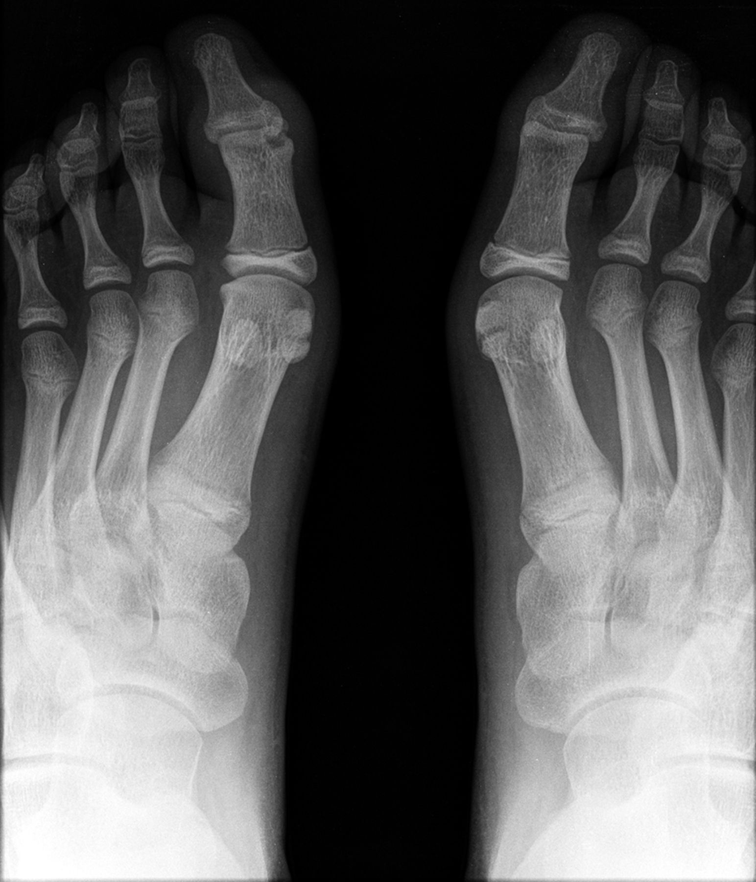
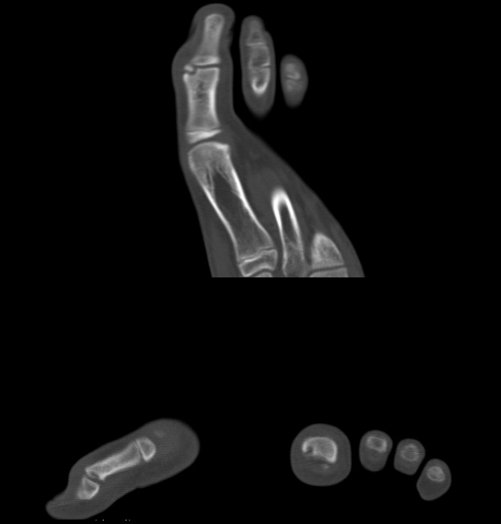
 Fig. 1
 Fig. 2
 Fig. 3
A fourteen-year-old Caucasian boy who was a regular practitioner of European football (soccer), playing as a goalkeeper, was referred to our orthopaedics consultation clinic; he reported sudden pain in the left hallux with athletic activity, particularly during contact with the ball. Physical examination revealed edema and tenderness at the dorsomedial aspect of the interphalangeal joint. The joint was stable and its range of motion was −5° of passive dorsiflexion to 30° of passive plantar flexion, equal to that on the contralateral foot, as measured roughly by goniometer. Active and passive maneuvers were painful. The other foot, his dominant one, sustained only local edema at the same spot. Complete blood-cell count, C-reactive protein, erythrocyte sedimentation rate (ESR), uric acid, and rheumatoid factor analyses were normal. A foot radiograph (Fig. 1) and computed tomographic (CT) scan (Fig. 2) revealed bilateral hyperlucent lesions on the medial side of the distal articular surface of the first phalanx of each hallux, with subchondral detachment on the left side.
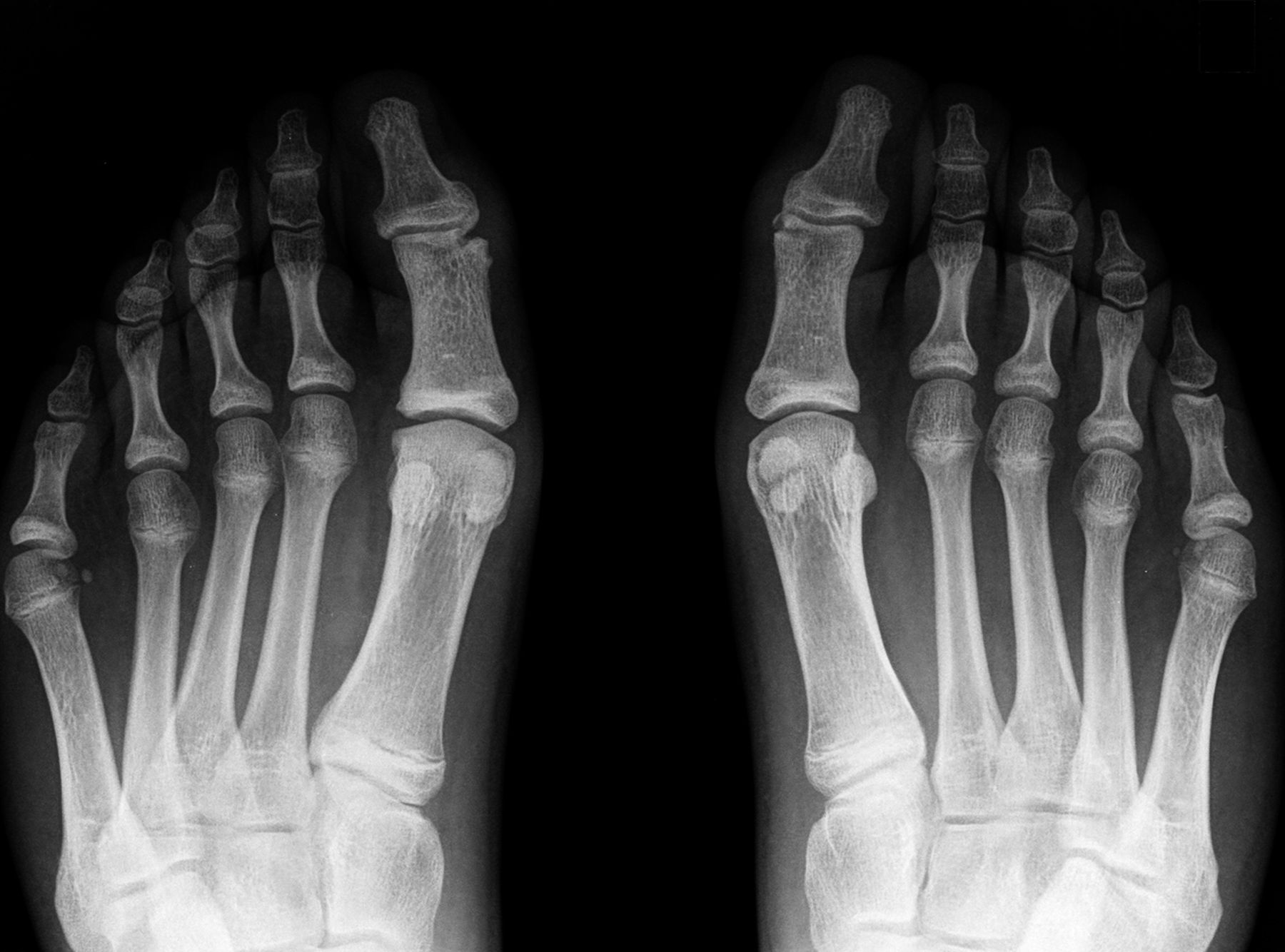
The patient was diagnosed with osteochondral fracture of the interphalangeal joint. After patient and parental consent, we performed surgery on the left hallux. We first performed a longitudinal sectioning of the ligament and capsule, thus allowing us to address the lesion without detachment of the medial collateral ligament. The lesion was subchondral, and we therefore performed resection (cartilage and bone), followed by tightening of the capsule through a straightforward transverse suture with the aid of a polymeric absorbable suture. The postoperative radiograph confirmed the removal of the lesion (Fig. 3). Complete weight-bearing was allowed after eight weeks, as we considered that, for the capsule to heal without lengthening, we should keep our patient from full weight-bearing on the interphalangeal joint for at least double the amount of time that soft tissue takes to heal. For logistical reasons, we evaluated the patient at about eight weeks postoperatively. At five months, he resumed sport-specific training. At the six-month follow-up, the lesion had resolved on a radiograph, and the patient reported no symptoms, resuming his activity as a goalkeeper without limitations. At the fourteen-month follow-up, his contralateral foot was still asymptomatic.
Proceed to Discussion >>Reference: Figueiredo SAL, Machado LML, Rodrigues J-MF, Sa AES. Osteochondral Lesions at the Interphalangeal Joint of the Hallux. JBJS Case Connect, 2015 Oct 28;5(4):e89.
There are several similarities between the case presented here and cases reported by Kinoshita et al. and Jones. Jones reported five lesions in the medial condyle of the first phalanx in military personnel, supposedly caused by repetitive trauma. In his observations, he noticed that all fragments were dorsomedial, had a fibrous attachment to the proximal phalanx, and were in connection with the collateral ligament. In our case, the fragment was loose from the first phalanx and distal to the ligament’s insertion, rather than at the insertion as was seen in the first case reported by Kinoshita et al. The lesion that we observed was positioned less dorsally, despite reports of symptoms on the dorsomedial aspect of the great toe. The treatment applied by Kinoshita et al. differed from ours in complexity, as, apart from lesion resection, it was followed by bone-graft interposition, fixated by either surgical wire or Kirschner wires. Despite treatment variations, the results were similar in the two cases reported by Kinoshita et al. and the current case, with patients returning to their previous activity (football or soccer) without restraints. Interestingly, our patient had radiographic evidence of lesions on both of his feet, and, although his right foot was dominant, he reported no symptoms on that foot. This suggests that not all lesions present with symptoms and, as such, their rarity may be due to underdiagnosis. It also suggests that a disruption of normal endochondral ossification may not be solely caused by recurrent minor trauma, as not only the dominant foot was affected.
Reference: Figueiredo SAL, Machado LML, Rodrigues J-MF, Sa AES. Osteochondral Lesions at the Interphalangeal Joint of the Hallux. JBJS Case Connect, 2015 Oct 28;5(4):e89.
Salter-Harris type-IV fracture of the proximal phalanx
Multiple epiphyseal dysplasia
Juvenile rheumatoid arthritis
Osteochondral fracture of the interphalangeal joint
Dislocation of the great toe