


 Fig. 1-A
 Fig. 1-B
 Fig. 2-A
 Fig. 2-B
 Fig. 3-A
 Fig. 3-B
 Fig. 4-A
 Fig. 4-B
 Fig. 5-A
 Fig. 5-B
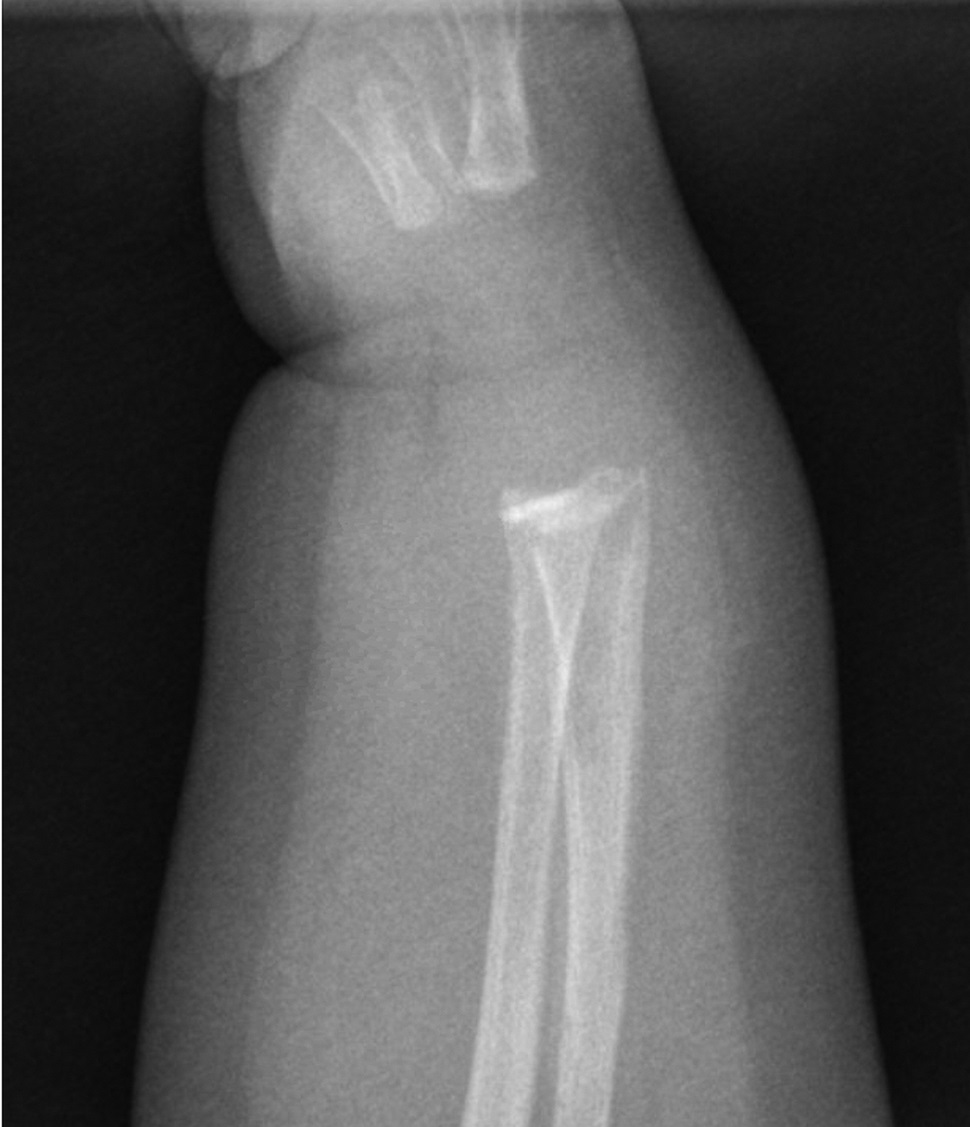
A 9-week-old girl was brought to the emergency room by her grandmother, who stated that the child was not moving her right arm. There was no known history of trauma. The patient had been afebrile and feeding regularly and had regular wet diapers and stooling. She was born full term without complications, was up to date on vaccinations, and had no other notable medical history. On examination, her vital signs were normal and she was in no distress. She was normocephalic with normal respiratory effort. Her right wrist was swollen and elicited a pain response with palpation. There were no other deformities of her extremities and no skin lesions.
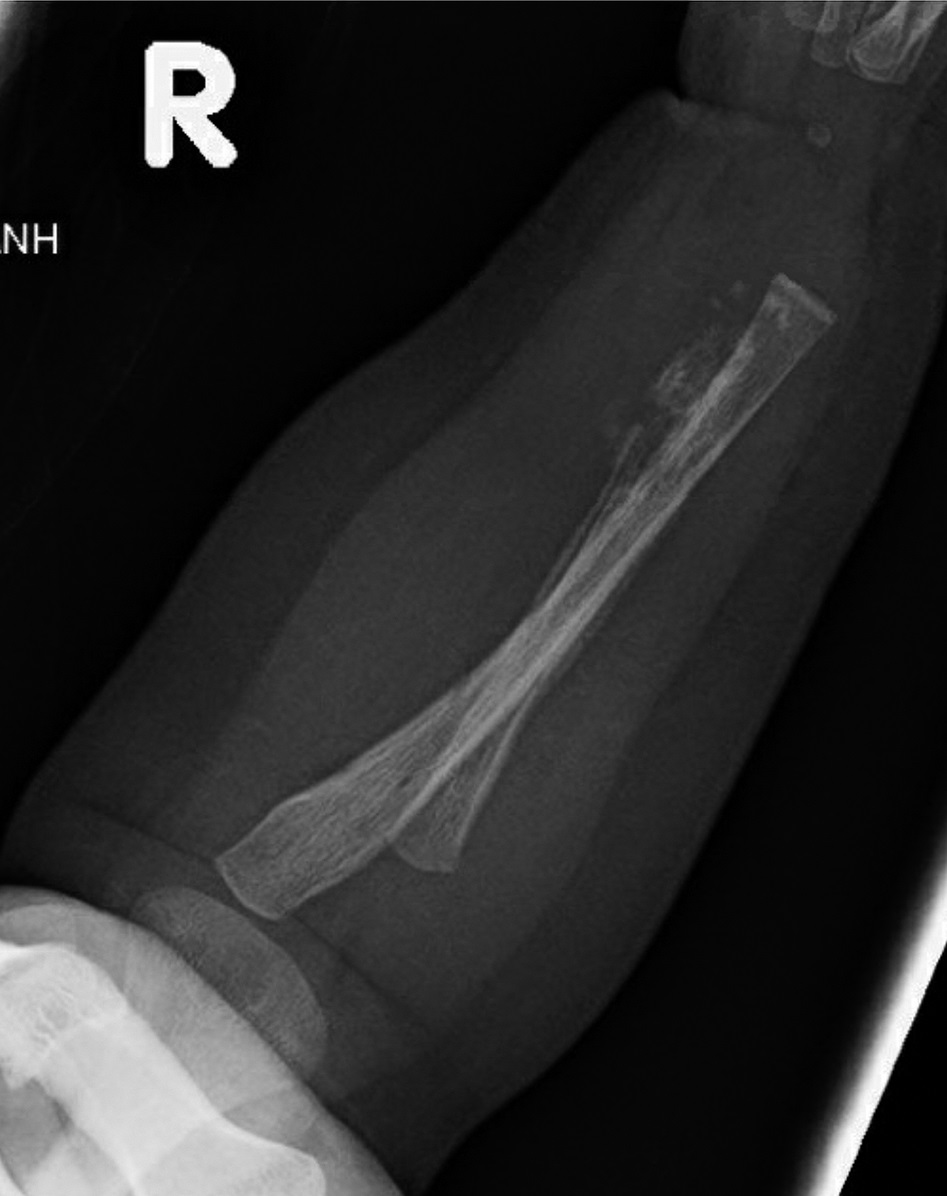
A radiograph of the right wrist was made and showed a subacute metaphyseal corner fracture of the distal part of the radius (Figs. 1-A and 1-B). Given the age and radiographic findings, a non-accidental trauma workup was initiated, per the institutional protocol. This included a computed tomographic (CT) scan of the head, abdomen, and pelvis, skeletal survey, blood work, and consultation to ophthalmology, general surgery, social work, Child Protective Services, and orthopaedics. The orthopaedic team evaluated the patient and placed the right arm in a soft immobilization, given the presumed subacute nature of the fracture. All other workup was negative, but, given the high suspicion for non-accidental trauma, the child was admitted and was subsequently discharged to foster care.
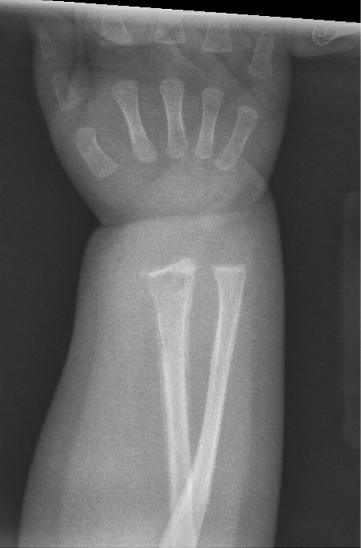
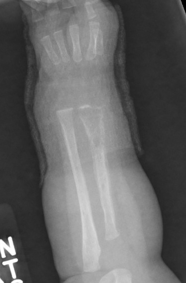
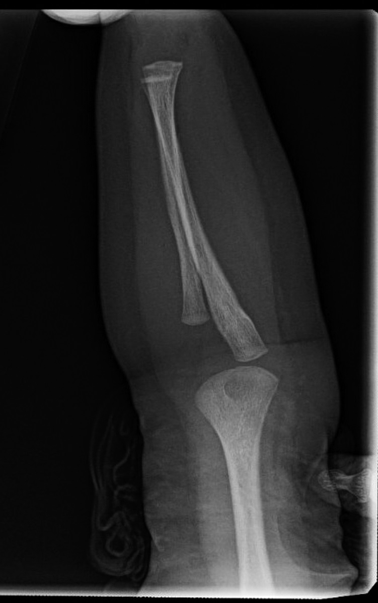
The patient was seen by a pediatrician 1 week later, and a repeat skeletal survey showed increased lucency of the right radius, interpreted as a “healing response to known fracture” (Figs. 2-A and 2-B). There was also a note of a “questionable periosteal reaction along the shaft of the right tibia” (Figs. 3-A and 3-B). Orthopaedics was not re-engaged after these findings, and the patient returned to the foster home.
Subsequently, the patient was tested and a rapid plasma reagin (RPR) screen was positive. The patient was brought to the Emergency Department, and a complete blood count (CBC), comprehensive metabolic panel, and cerebrospinal fluid (CSF) testing were obtained. The results were positive for transaminitis, hyperkalemia, and anemia. The CSF test was positive for venereal disease research laboratory (VDRL) testing. The patient was admitted to the Pediatrics Department, an infectious disease consultation was obtained, and the patient was administered intravenous benzathine penicillin G (BPG).
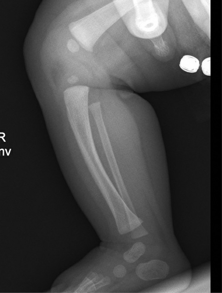
Repeat forearm radiographs were made 1 week later; these showed progressive lucency of the radius with osteolysis and cortical destruction (Figs. 4-A and 4-B). In addition, there were lesions noted in the left radius and ulna as well (Figs. 5-A and 5-B). The forearm was not further immobilized, and the patient was followed clinically by our orthopaedics team. The patient remained in the hospital to complete her course of antibiotics.
Clinical features, radiographic findings, and the results of RPR and VDRL testing led to the diagnosis of congenital syphilis. The Office of Public Health confirmed that the biological mother had tested positive for syphilis at the time of delivery and was undergoing treatment, although, reportedly, this finding in the mother was not documented. At the last follow-up, the parents had been cleared of non-accidental trauma and the patient was doing well.
Proceed to Discussion >>Reference: Hariharan AR, Schindelar L, Nichols LRB, Kruse RW. A metaphyseal corner fracture that wasn’t. A case report of osteitis from congenital syphilis. JBJS Case Connect. 2020;10(1):e0557.
When evaluating a neonate with radiographic evidence of a fracture or lytic lesion, diagnoses other than non-accidental trauma must be kept in mind. Although a rare condition, congenital syphilis must remain on the differential for any lytic bone lesion in a young infant.
Infants with congenital syphilis may present with typical signs and symptoms or may be asymptomatic. The clinical findings may become clearer over several months of life, but the array of these findings is vast. Signs of early congenital syphilis include skin rash, hepatosplenomegaly, rhinitis (“snuffles”), condyloma lata, cranial nerve palsies, and seizures. Our patient displayed none of these signs. Laboratory findings may demonstrate anemia, transaminitis, and hyperbilirubinemia. Radiographic evidence may reveal pneumonia alba, periostitis, osteochondritis, and Wimberger sign (destruction of the proximal tibial metaphysis). The skeletal abnormalities often involve the long bones, the ribs, and the cranium. The bone lesions may be painful and cause pseudoparalysis (pseudoparalysis of Parrot). Infected infants may display several sequelae of the disease including cerebral palsy, hydrocephalus, deafness, glaucoma, myocarditis, nephrotic syndrome, and musculoskeletal deformities such as frontal bossing, saber shins, and sternoclavicular thickening.
Diagnosis of congenital syphilis can be difficult when classic signs and symptoms are absent or the maternal history is unclear. Congenital syphilis should be suspected when the mother has untreated syphilis at delivery or in a child who has evidence of congenital syphilis on examination or radiographs. In suspected cases of congenital syphilis, a complete diagnostic evaluation is warranted, including serologic testing, long bone radiographs, CBC, and a lumbar puncture.
First, congenital syphilis can be confirmed in an infant when Treponema pallidum is identified by darkfield microscopy or polymerase chain reaction in body fluid or tissue specimens. However, all children with suspected disease should have serologic testing. Second, long bone radiographs should be made. They are abnormal in up to 65% of babies with congenital syphilis symptoms but can be as low as 5% when asymptomatic. Therefore, the results of radiographs in many infected children do not reliably lead to a diagnosis, but abnormalities on radiographs may provide the earliest evidence of infection, even before serological results. Third, the diagnosis of neurosyphilis is difficult to establish because many children with congenital syphilis do not manifest any neurological symptoms. An elevated CSF cell count (>18 to 25 cells/mm3), an elevated protein level (>0.15 g/L), and a positive VDRL result are suggestive of neurosyphilis but have low sensitivity and specificity. Nevertheless, if clinical or laboratory testing is suggestive of congenital syphilis, then therapy effective against neurosyphilis is warranted. Finally, additional studies such as chest radiographs, liver function tests, abdominal ultrasound, neuroimaging, and auditory and ophthalmologic examination may be warranted.
Children with proven disease or with high likelihood of disease include those with a serum quantitative nontreponemal serologic titer 4 times higher than that of the mother, a positive darkfield test, or an abnormal physical examination or radiographic finding consistent with congenital syphilis. Aqueous BPG is the preferred choice of treatment in these children. Children younger than 4 weeks of age are treated with BPG 50,000 units/kg intravenously every 12 hours for 7 days, followed by every 8 hours for 14 days. For infants older than 4 weeks of age, the dose is 50,000 units/kg every 6 hours intravenously for 14 days. The increased frequency is to cover potential neurosyphilis. During the first 24 hours of treatment, some patients may experience a Jarisch-Herxheimer reaction, which is assumed to be due to an inflammatory reaction to the dying spirochetes. This presents as fever, chills, hypotension, and tachycardia and is managed with supportive care.
Children with congenital syphilis should have serial VDRL/RPR serology testing, and these titers should become nonreactive by 12 months after treatment. Those with abnormal CSF findings on initial diagnosis should have a repeat lumbar puncture 6 months after treatment. If appropriate and reliable follow-up of an infant cannot be ascertained, the child should be kept in the hospital for the full course of therapy. Most importantly, a multidisciplinary approach and a regular follow-up are critical for the care of these children.
The incidence of congenital syphilis is increasing and can often be difficult to diagnose, particularly when the maternal history is unclear. As the orthopaedic provider, it is important to keep this differential in mind when evaluating a patient with lytic long bone lesions or questionable metaphyseal injuries. Congenital syphilis is a treatable condition that may result in death if missed at presentation, and early diagnosis may prevent or ameliorate the sequelae of the disease.
Reference: Hariharan AR, Schindelar L, Nichols LRB, Kruse RW. A metaphyseal corner fracture that wasn’t. A case report of osteitis from congenital syphilis. JBJS Case Connect. 2020;10(1):e0557.
What is the diagnosis?
Chronic recurrent multifocal osteomyelitis
Probable non-accidental trauma
Hepatitis with biologic false-positive RPR and VDRL
Congenital syphilis
Multifocal Langerhans cell histiocytosis (Letterer-Siwe disease)