

 Fig. 1
 Fig. 2
 Fig. 3
 Fig. 4
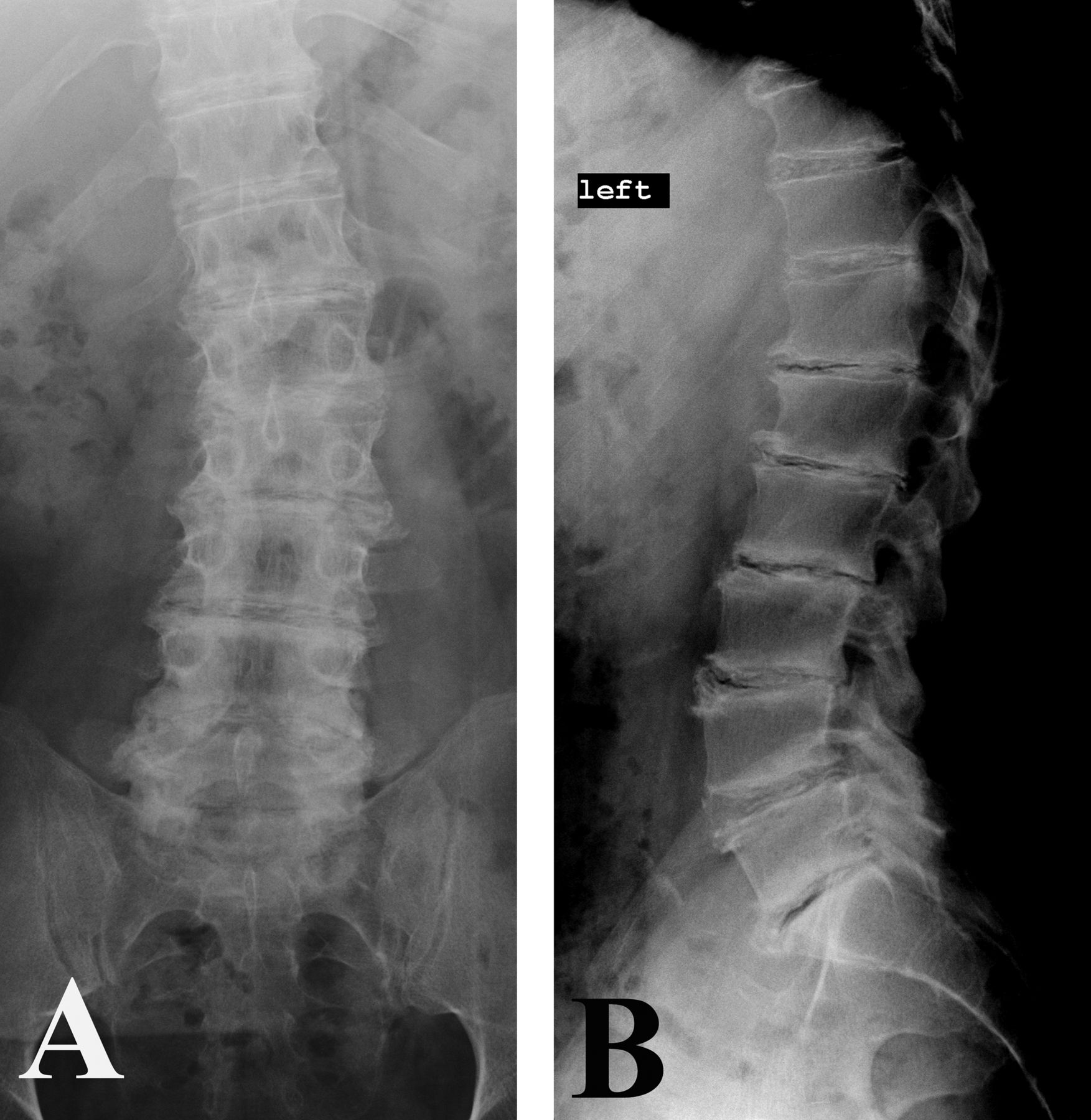
A 39-year-old man presented with low back pain. Imaging demonstrated lumbar scoliosis, extensive irregularities of end-plate vertebral margins, and advanced Thompson Grade-V disc degeneration (Fig. 1). Six months later, the patient presented with lower-extremity radiculopathy. MRI studies were performed, and a herniated L2-L3 disc was identified (Fig. 2). End-plate resorption and irregular disc margins were very prominent features.
Minimally invasive surgical disc excision was performed for extrusion of the herniated disc at L2-L3 on the left. Radicular pain resolved after surgery. At the 2-year follow-up, he had diffuse neck and thoracolumbar spine pain.
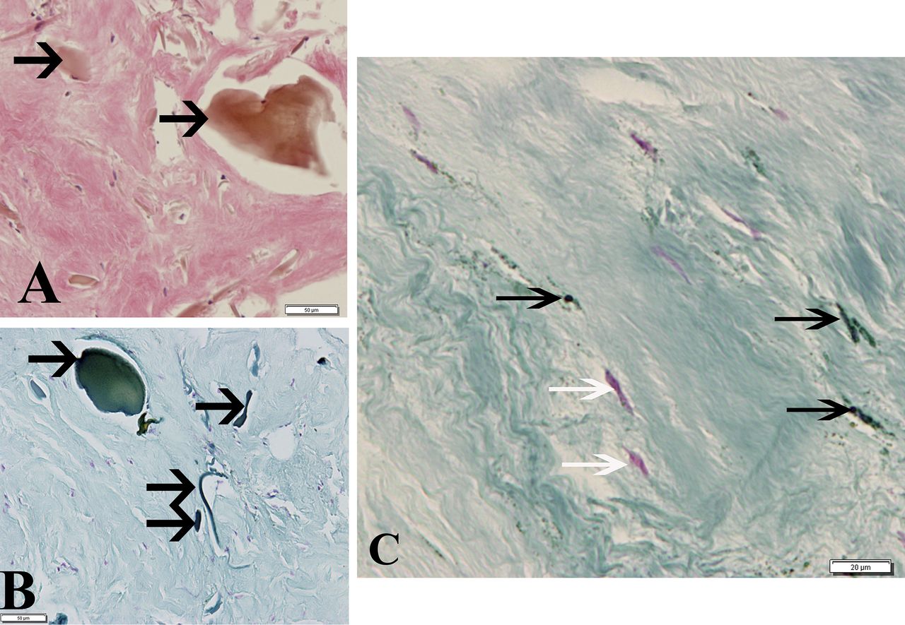
The excised disc fragment was noted to be black, with severe degeneration. Routine light microscopy and the use of special stains are shown in Figure 3.
Disc cells and extracellular matrix were also examined in greater detail using transmission electron microscopy (Fig. 4).
The tissue was processed for routine and undecalcified processing as well as ultrastructural examination. Routine light microscopy revealed a sparse cell distribution in the anulus tissue and the presence of large pigment granules in the extracellular matrix. Specialized staining with Schmorl reagent provided a sensitive method for detection of ochronotic pigment as previously shown by Preston et al. and Ranganath et al. Within the disc tissue studied, the pigment inclusions appeared larger than those previously reported within joint tissues. These findings confirm the diagnosis of ochronosis (alkaptonuria [AKU]).
Proceed to Discussion >>Reference: Gruber HE, Hanley EN Jr. Alkaptonuria-associated changes in disc degeneration: a case report. JBJS Case Connect. 2015 Nov 11;5(4):e96.
The disease progression and natural history of AKU are still poorly understood; Ranganath et al. recently noted that patients appear to become symptomatic at approximately 30 years of age, at which time the slowly progressing ochronosis causes overt pathology, and this is followed by a period with marked increase in pathology at 50 to 60 years. The Alkaptonuria Severity Score Index (AKUSSI) includes scales developed to assess both joint-pain issues (joint pain, need for arthroscopy, and need for joint replacement) and clinical spinal features. Muscle, ligament, and tendon ruptures are additional disease-related events. Nonskeletal events include hearing impairment, stroke, and aortic sclerosis.
Fusion of vertebrae was a finding reported by Galdston et al. As AKU progresses, back pain and stiffness as well as loss of normal sagittal alignment occur. Disc herniation may be an initial presenting feature in the lumbar spine.
In the case presented in this report, end-plate resorption and irregular disc margins were prominent features. End-plate changes were previously noted in the literature, but are sometimes difficult to interpret with poor-resolution older radiographs. Hamdulay et al. noted diffuse, type-2 Modic end-plate changes in their patient, and Palazzi et al. identified changes in the thoracic end plates. MRI findings in AKU have not been well documented to date. It is interesting to note that research examining failed articular joints of alkaptonuric patients shows aggressive osteoclastic resorption of highly pigmented cartilage matrix regions in the subchondral plate. It is possible that similar heightened osteoclastic activity is underway in the end-plate cartilage of AKU vertebrae.
The diagnosis of AKU should be suspected when articular cartilage in any joint or fibrocartilage of the intervertebral disc has a black appearance during an operation. Only a few studies on disc histology in AKU have illustrated small pigmented deposits and larger deposits. To our knowledge, the ultrastructural findings reported here represent the first description of large AKU deposits in the human disc extracellular matrix and within anulus cells themselves. We identified small intracellular shards and electron-dense pigment present on the surface of collagen fibers—features very similar to findings by Taylor et al. in the ligamentous joint capsule of the hip of an alkaptonuric patient undergoing hip replacement.
The accumulated masses of ochronotic pigment granules may be quite important within the disc, where structural integrity of the matrix is biologically critical. These masses may increase with aging of the alkaptonuric patient, and they not only may disrupt the extracellular matrix (as previously recognized in cartilage containing crystal deposits and large crystal masses in the disc) but also could potentially contribute to degenerative matrix changes via elevated matrix metalloproteinase expression and altered disc-cell gene expression. Since failure of the structural integrity of the disc can result in an anular tear with subsequent disc herniation, the mechanisms of ochronotic pigment formation and the relation of these pigments to disc degeneration and vertebral end-plate resorption merit further investigation.
Reference: Gruber HE, Hanley EN Jr. Alkaptonuria-associated changes in disc degeneration: a case report. JBJS Case Connect. 2015 Nov 11;5(4):e96.
What is the diagnosis?
Adverse local tissue reaction to metal debris
Gaucher disease
Pompe disease (glycogen storage disease type II)
McArdle disease (glycogen storage disease type V)
Ochronosis (alkaptonuria)