A 31-Year-Old Man with Knee Pain Following Gym Exercise
December 7, 2022
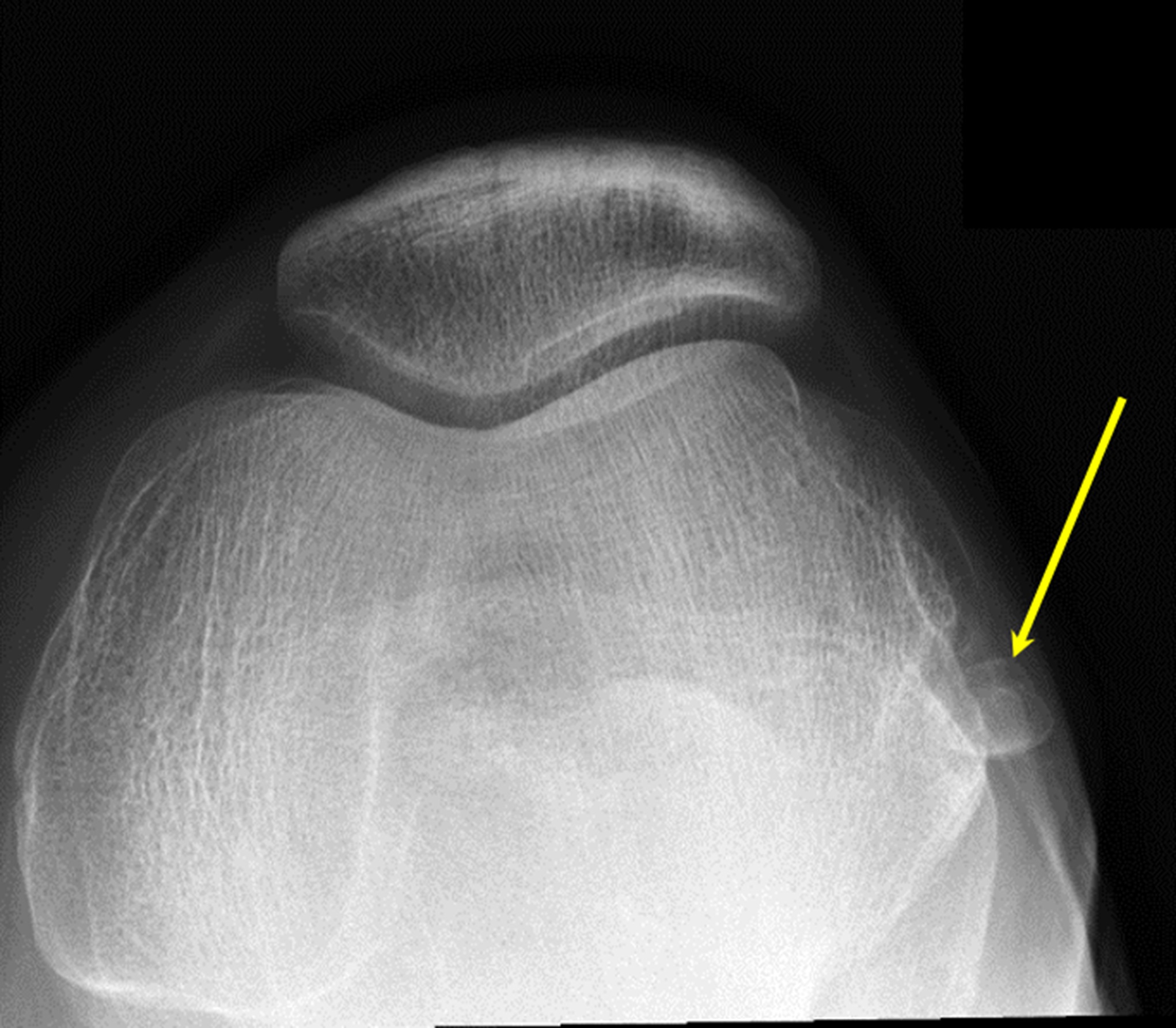
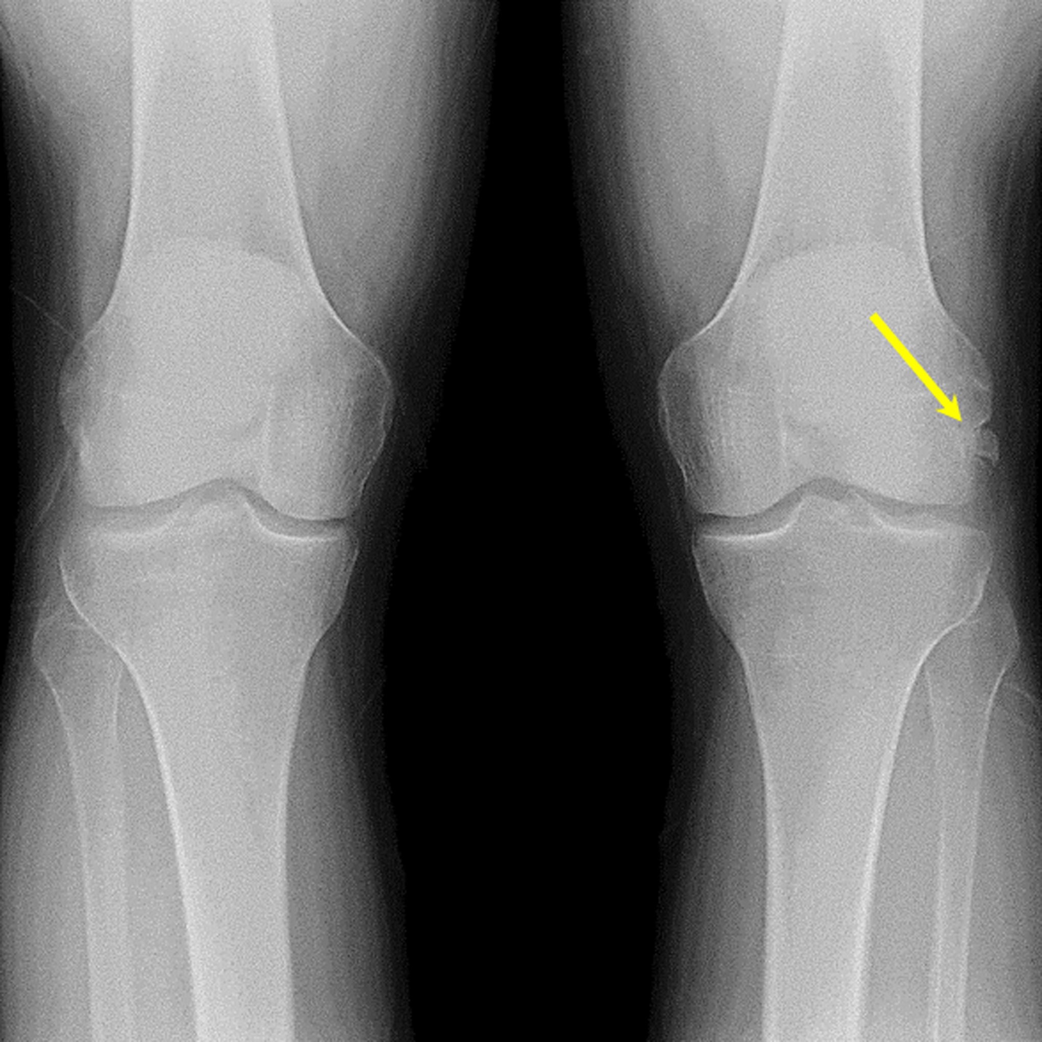
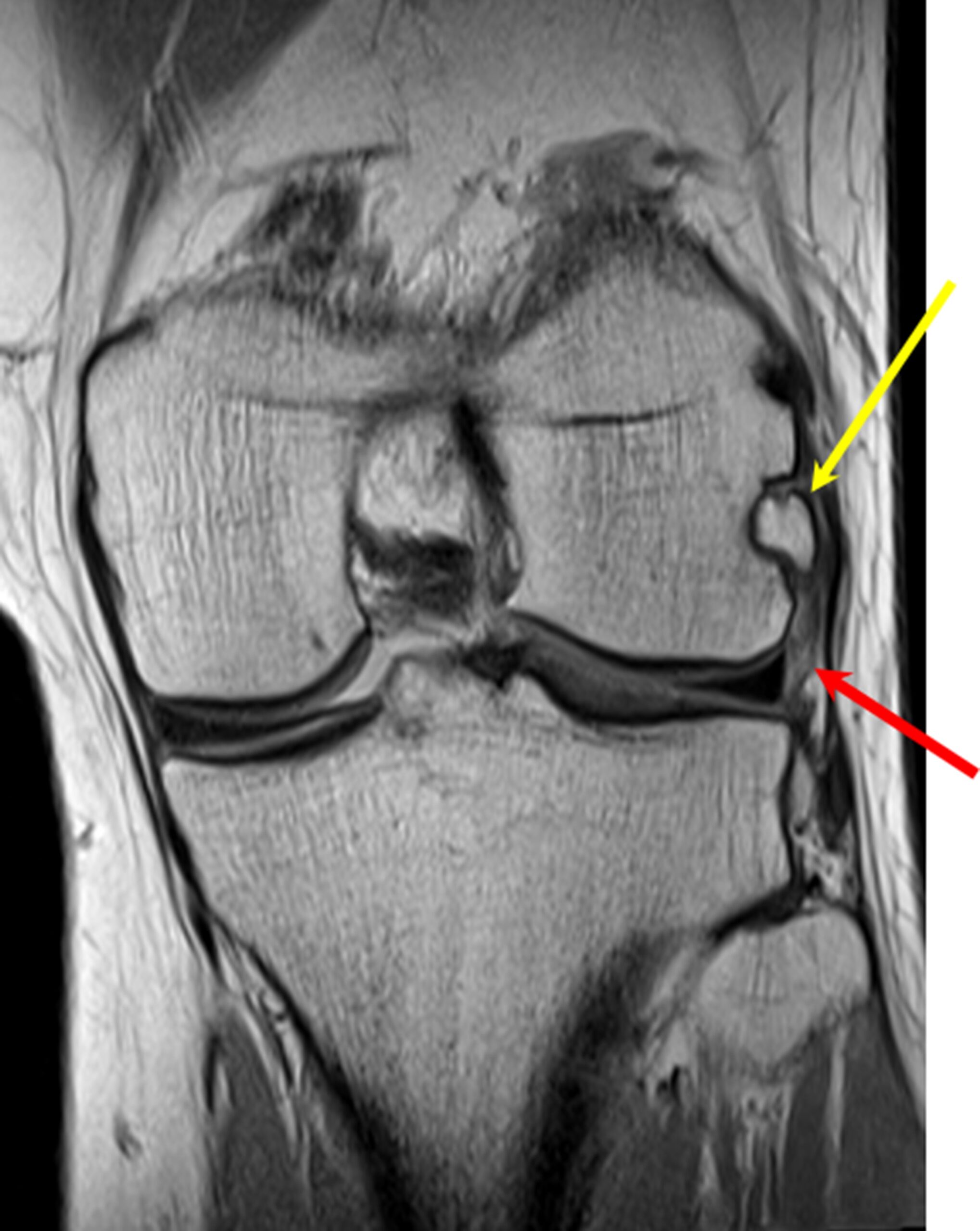
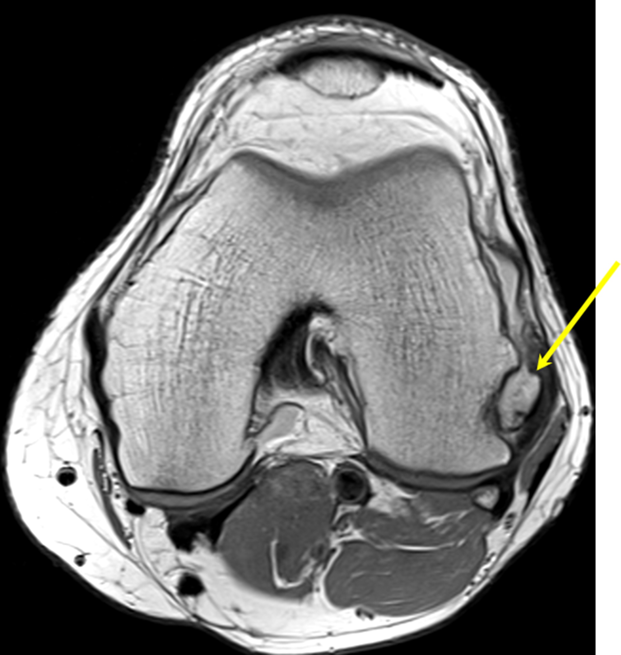
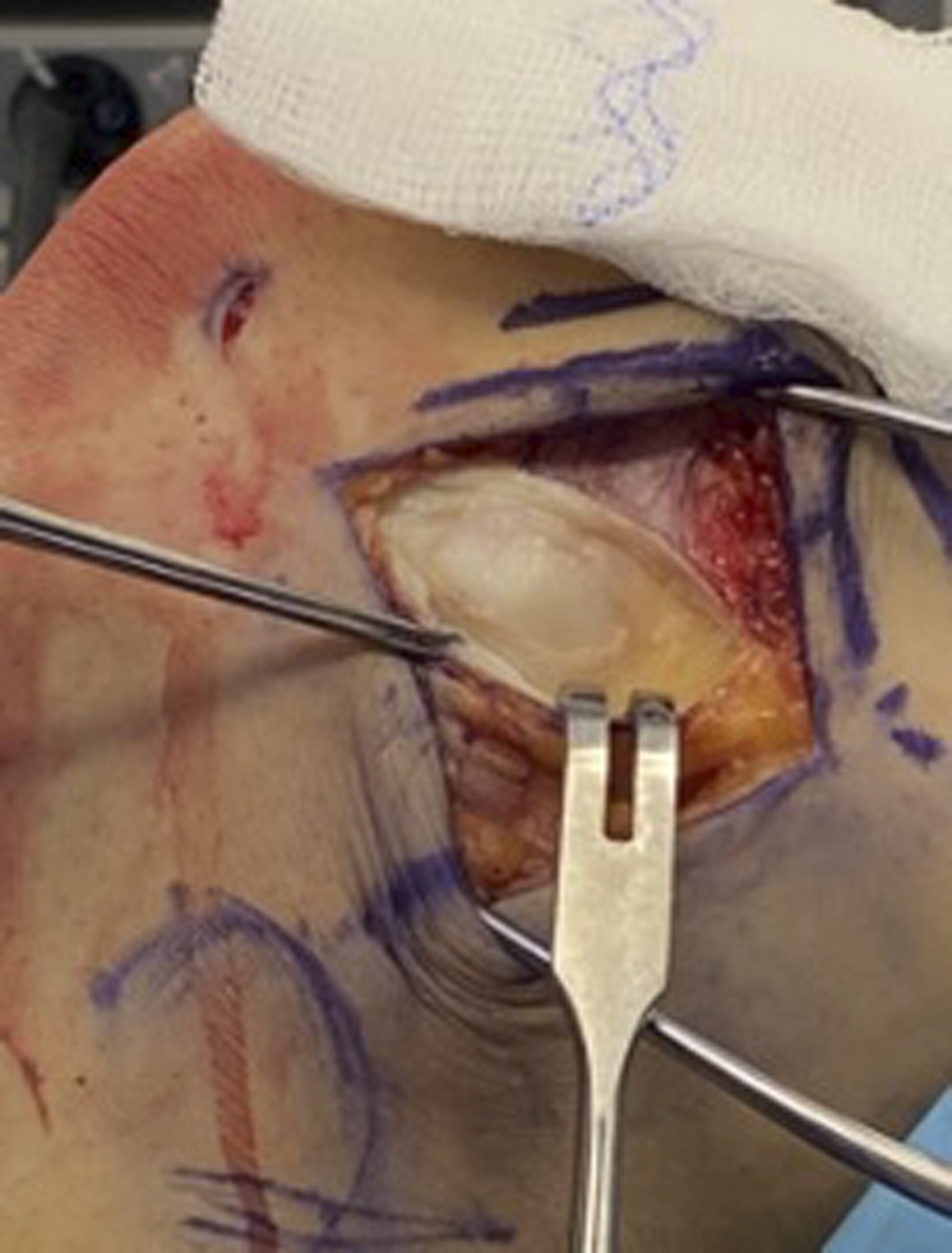
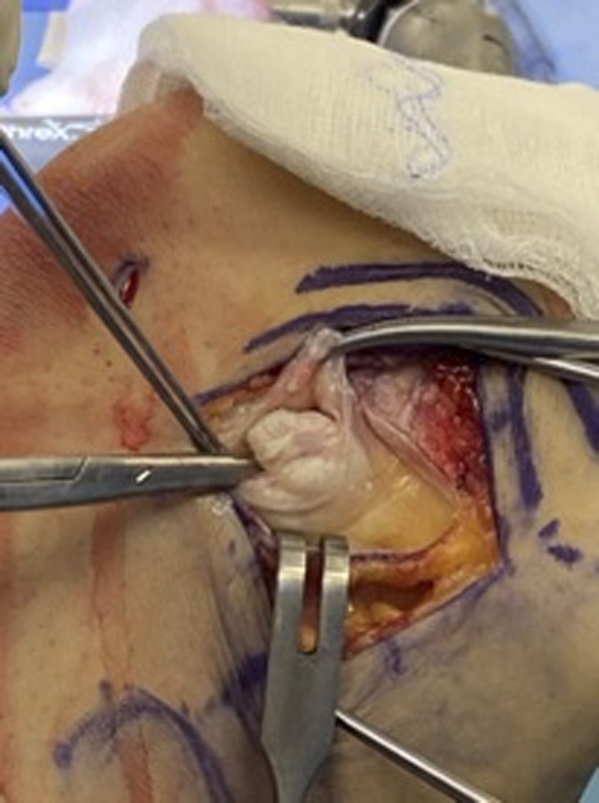
 Fig. 1-A
 Fig. 1-B
 Fig. 2-A
 Fig. 2-B
 Fig. 3-A
 Fig 3-B
A 31-year-old man presented with left knee pain and knee locking that occurred only with flexion during squatting exercises at the gym. Eighteen years earlier, he had sustained a blow to the outstretched knee in a lacrosse event. He had been managed with immobilization followed by 3 months of physical therapy.
On physical examination at presentation, a nodule was noted to be mobile with the knee in the flexed position. The nodule would move distally and then lock the knee, with maximum flexion of the knee. To unlock, the knee had to be flexed and the nodule would have to be manipulated in the proximal and anterior directions, thus allowing for full mobility. The physical examination of the knee was otherwise negative with intact ligaments.
Imaging included radiographs that demonstrated a 1.2 × 1.6 cm nodule lateral to the left femoral condyle (Figs. 1-A and 1-B). Follow-up magnetic resonance imaging (MRI) scans showed the nodule in the popliteus sulcus, thinning of the popliteus tendon, and a trace joint effusion (Figs. 2-A and 2-B). The decision was made to excise the nodule.
Arthroscopy of the knee was performed with standard anteromedial and anterolateral portals. Diagnostic arthroscopy demonstrated intact chondral surfaces with no meniscal abnormalities. The cruciate ligaments were intact. The lateral gutter was examined, and the nodule was found to be extra-articular. It was decided to proceed with open excision.
Next, an incision was made centered over the lateral epicondyle. The iliotibial band was split, and the nodule was identified and delivered bluntly (Figs. 3-A and 3-B). The wound was closed in a layered fashion.
The combination of imaging and intraoperative findings led to the diagnosis of a chronic popliteus avulsion fracture with atrophy of the popliteus tendon.
Immediately postoperatively, the patient had full extension and flexion of the knee without locking. The patient was discharged home the same day and was instructed to bear weight as tolerated. Within 3 days, he was walking with a normal gait. He performed physical therapy 2 times per week for a total of 6 weeks, followed by an independent strength training program. At 6 months after the surgical procedure, he had no pain or locking and full range of motion.
Proceed to Discussion >>Reference: Blackwell C, Selley R, Taber CE, Benitez CL, Marx RG. Chronic popliteus tendon avulsion fracture with chronic knee pain and locking: a case report. JBJS Case Connect. 2022 Jan 26;12(1).e21.00477.
This is the second documented case of the surgical treatment of a chronic popliteus avulsion fracture. In the first documented case, nonoperative management ultimately led to a valgus deformity secondary to the formation of a lateral physeal bone bridge, whereas, in this case, conservative treatment ultimately resulted in a locked knee. This case raises the question as to whether nonoperative management of an acute avulsion fracture is the best approach. Furthermore, several case reports have noted the success of early intervention in acute popliteus avulsion fractures with successful outcomes.
Several authors have argued that nonoperative management of isolated popliteus tendon avulsion fractures is indicated when there is no ligament injury. Although long-term outcomes of conservative management are not well studied, it is clear that, in the short term, these patients can return to full activities within a few months and can avoid the recovery and risks associated with a surgical procedure. Although a proportion of patients treated nonoperatively will ultimately require surgical intervention, the majority will likely remain asymptomatic, furthering the case for initial conservative management. Furthermore, this injury to the popliteus tendon could have long-term implications, should the patient require a total knee arthroplasty at a later date.
It is difficult to know which patients would be better treated with an early surgical procedure. One useful tool, which has not been documented in any of the previous case reports, would be a computed tomographic (CT) scan of the knee. A CT scan could assist in quantifying the size and morphology of the avulsed fragment and the degree of the injury, which could direct treatment options to help to determine the healing potential of the fragment. In our case, the bone fragment went on to a nonunion, which ultimately necessitated surgical intervention. If nonoperative treatment is performed, the patient should undergo close follow-up to determine whether a surgical procedure is indicated on a delayed basis either for symptoms or for deformity secondary to a growth arrest.
Reference: Blackwell C, Selley R, Taber CE, Benitez CL, Marx RG. Chronic popliteus tendon avulsion fracture with chronic knee pain and locking: a case report. JBJS Case Connect. 2022 Jan 26;12(1).e21.00477.
What is the diagnosis?
Periosteal osteoid osteoma
Chronic popliteus avulsion fracture
Myositis ossificans
Synovial chondromatosis
Osteochondroma