
 Fig. 1
 Fig. 2
 Fig. 3
 Fig. 2
 Fig. 3
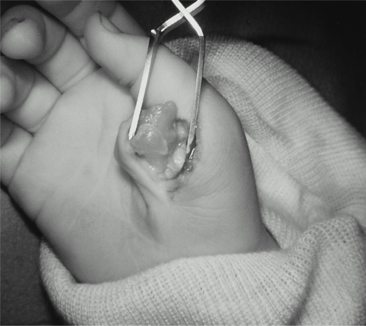
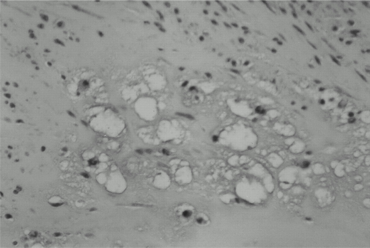
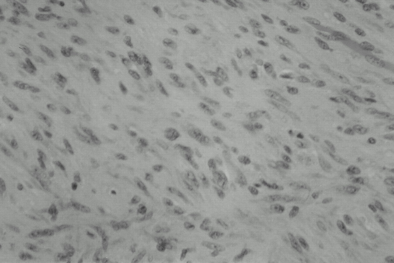
A two-year-old girl with no history of any medical problems presented with a mass on the volar radial aspect of the right thumb at the level of the metacarpophalangeal joint and extending onto the thenar eminence. The family had just recently noticed the mass. We observed normal motion of all of the thumb joints, with normal active function of the extensor pollicis longus and the flexor pollicis longus muscles. There was no triggering, and the patient appeared to use the hand normally. The mass was mobile and did not seem to be painful. Sensory testing could not be done because of the child’s age. Radiographs did not demonstrate any osseous involvement, but there was calcification in the soft-tissue mass. At surgery, a flesh-colored lobulated mass measuring 1.7 × 1.0 × 0.9 cm was found (Fig. 1). The radial digital nerve of the thumb was closely adherent to the mass on its inferior surface. The mass was dissected out with use of 3.5-times loupe magnification, and the radial digital nerve was not compromised. Microscopic sections revealed a spindle-cell neoplasm with one mitosis per ten high-powered fields. There was no atypia. Histological sections stained with hematoxylin and eosin revealed a pattern of interlacing bundles of spindle cells (Fig. 2), with scattered foci of chondroid differentiation (Fig. 3). Sections with special staining, including S-100 protein, smooth-muscle actin, and Masson trichrome, were reviewed. Diffuse background staining with S-100 protein was noted, but this was thought to be nonspecific; there was definite staining with smooth-muscle actin; and smooth-muscle elements were noted on Masson trichrome staining.
A pathological diagnosis of calcifying aponeurotic fibroma was made. Postoperatively, the patient did well, and the lesion had not recurred at the time of this writing
Proceed to Discussion >>Reference: DeSimone RS, Zielinski CJ. Calcifying aponeurotic fibroma of the hand. A case report. J Bone Joint Surg Am. 2001 Apr;83(4):586-8.
In 1953, Keasbey first described calcifying juvenile aponeurotic fibroma in four children. Keasbey and Fanselau changed the term to aponeurotic fibroma in 1961, after it became apparent that the condition was not limited to children. Lichtenstein and Goldman named the same entity cartilage analogue of fibromatosis in 1964. In 1973, Iwasaki and Enjoji used the term calcifying aponeurotic fibroma, which is the current usage endorsed by Enzinger and Weiss. Most textbooks devote only a cursory paragraph to this condition. In 1977, Wallace and Fitzmorris reviewed the literature and noted several cases of faulty diagnosis; the most common misdiagnoses were sarcoma, fibrosarcoma, and pseudosarcoma. These misdiagnoses led to amputation and other radical surgery, resulting in decreased function. The usual presentation is a painless mass. However, Allen and Enzinger noted two cases with mild pain and tenderness, and Keller and Baez-Giangreco reported three cases in which major pain was a presenting complaint. The mass rarely measures more than 3 cm in diameter. To our knowledge, it has never been reported as being adherent to the skin or associated with contractures. It usually appears as a poorly encapsulated mass of moderately cellular, dense fibrous tissue, which infiltrates fat, voluntary muscle, and collagen bundles or surrounding nerves. Radiographs may show a soft-tissue mass with no associated osseous lesions and a fine stippling of focal calcification. The tumor was initially described in the palms, fingers, and soles of the feet of children. Enzinger and Weiss reported that 77% of patients had a tumor in the hand and 13% had a tumor in the foot (numbers not given). They also reported that 70% of the tumors were in boys. The tumor has since been reported in the wrist, forearm, elbow, upper arm, neck, abdominal wall, lumbar paravertebral area, leg, and ankle. The majority of these tumors appear in children in the first and second decades of life. The median age at the time of diagnosis is twelve years. The condition has been noted from birth to sixty-four years of age. There is no evidence of any increased familial prevalence. Keasbey and Fanselau described a distinct cell type containing large, darkly staining, plump, oval nuclei, with no discernible cytoplasm, oriented in one direction in a sheet (Fig. 2). Lichtenstein and Goldman stressed that the pattern was nodular and that chondroid differentiation was the unique feature of this entity (Fig. 3). The term aponeurotic has been used because there is a subtle transition from fibrous connective tissue to fibrocartilage as tendons, aponeuroses, and ligaments insert into bone through Sharpey fibers, and the location of many of these tumors suggests an aponeurotic origin. All authors have reported areas of calcium deposition, which do not occur in areas of degeneration. Two phases in the tumor’s development have been described: an initial phase, more common in infants and young children, in which the tumor grows diffusely, and a late phase, in which the lesion is more compact and nodular. Calcification and chondroid differentiation are more common in tumors removed from older children and young adults. The clinical differential diagnosis includes fibrous hamartoma of infancy, diffuse infantile fibromatosis, Dupuytren disease, and chondroma of soft parts. Postoperatively, 50% of the tumors recur locally, but we know of no report of malignant transformation or metastasis. The masses rarely cause complications, and functional deficits are usually iatrogenic rather than the result of tumor growth. Treatment consists of excision to facilitate the diagnosis and to remove the mass. Reexcision may be performed if necessary, but observation of a recurrent lesion is certainly an option if there is no functional impairment.
Reference: DeSimone RS, Zielinski CJ. Calcifying aponeurotic fibroma of the hand. A case report. J Bone Joint Surg Am. 2001 Apr;83(4):586-8.

Calcifying aponeurotic fibroma
Fibrous hamartoma of infancy
Infantile fibromatosis
Fibrosarcoma
Chondroma of soft tissue