A 3-Year-Old Boy with Swelling of the Left Forearm Following an Accident
October 5, 2016
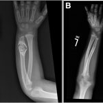
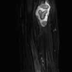
A 3-year-old boy presented with mild swelling in the left forearm after sustaining a fall on a trampoline. Initial radiographic examination revealed a pathologic fracture through a lesion in the distal aspect of the left ulna. Radiographs demonstrated an osteolytic lesion with sclerotic margins, mild expansion, and central calcification and then the results at the 18-month follow-up after treatment (Figs. 1-A and 1-B). MRI (magnetic resonance imaging) performed with and without contrast demonstrated a contrast-enhancing lesion in the distal diaphysis of the ulna with mixed signal intensity on T2 weighting (Fig. 2).
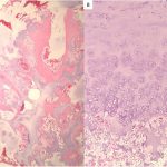
The patient underwent open biopsy, but a definitive diagnosis could not be made on the basis of this single biopsy sample (Fig. 3).
In light of this diagnostic uncertainty and the history of bone destruction to the point of pathologic fracture, the decision was made to perform definitive operative treatment. Seven days following the open biopsy, the lesion was resected by means of intralesional curettage and high-speed burring, then packed with crushed cancellous allograft.
Histologic examination of the resected lesion showed findings identical to those of the initial biopsy while additionally showing that the lesion had extended through the cortex and underneath the periosteum.
The combination of histologic findings and the intraoperative observations indicated that the best diagnosis is enchondroma protuberans. Following surgery, the patient was treated nonoperatively in a long arm cast for 4 weeks. The patient had an uneventful recovery, and follow-up at 18 months showed a dramatic decrease in the size of the lesion and restoration of a normal pattern of trabeculation (Fig. 1-B).
Proceed to Discussion >>Reference: Lasater P, Steensma MR, Patthanacharoenphon C, Davis MM. Enchondroma protuberans of the ulna in a pediatric patient: A case report. JBJS Case Connect. 2016 Jul 13;6(3):e54.
Radiographically, enchondroma protuberans appears as an intramedullary osteolytic lesion with a sclerotic margin. It may also be accompanied by well-defined cortical and soft-tissue expansion with a cortical defect. Radiographs may be all that is necessary to diagnose enchondroma protuberans. If MRI is utilized, the lesion will show low signal intensity on T1-weighted images and high signal intensity on T2-weighted and STIR (short tau inversion recovery) images.
Histologically, enchondroma protuberans displays columns of chondrocytes. The chondrocytes are uniform in appearance and are typically situated in lacunae within a myxoid matrix. Intermittent areas of calcification and sporadic foci of ossification are occasionally seen within this conglomerate.
Enchondroma protuberans is thought to originate from cartilaginous material inside the medullary canal of a long bone. Protrusion of the lesion through cortical bone is a characteristic of enchondroma protuberans that differentiates it from enchondroma. Enchondroma protuberans can further enlarge outside the cortex and lead to an expansile mass that may include calcifications or ossification. The most common location for enchondroma protuberans, as for enchondroma, is within the bones of the hand.
Enchondroma protuberans should also be differentiated from osteochondroma. Osteochondroma is an exophytic lesion that typically has a cartilaginous cap on the outer surface of a trabeculated osseous lesion, but no cartilaginous material is found within the medullary canal of the long bone or the base of the pedunculated lesion. Enchondroma protuberans exhibits cartilaginous material within the medullary canal of the bone and expansion of the mass beyond the cortex.
Periosteal chondroma may also mimic enchondroma protuberans. The distinction between the two is somewhat arbitrary, but MRI or histology shows less intramedullary involvement by a periosteal chondroma compared with an enchondroma protuberans. Chondrosarcoma can also resemble enchondroma protuberans. Discrimination between them is made with histologic examination. Enchondroma protuberans shows benign cartilage cells that are well differentiated. In contrast, chondrosarcoma shows features of pleomorphism, binucleation, increased cellularity, and occasional atypia. Chondrosarcoma often presents with pain, while a benign cartilage lesion is rarely painful unless complicated by fracture or trauma.
The patient in the present case had good clinical results following intralesional curettage. At the time of final follow-up, there has been healing of the pathologic fracture and no recurrence of the lesion. Intralesional curettage and grafting performed for a pathologic fracture through an existing enchondroma protuberans in the distal aspect of the ulna of a pediatric patient appears to yield satisfactory clinical results on the basis of our isolated experience with this rare lesion.
To our knowledge, this is the only case report documenting enchondroma protuberans in the ulna of a pediatric patient and only 1 other case report of enchondroma protuberans in the ulna has been documented, in a 20-year-old woman.
Reference: Lasater P, Steensma MR, Patthanacharoenphon C, Davis MM. Enchondroma protuberans of the ulna in a pediatric patient: A case report. JBJS Case Connect. 2016 Jul 13;6(3):e54.
What is the diagnosis?
Enchondroma
Periosteal chondroma
Low-grade chondrosarcoma
Osteochondroma
Enchondroma protuberans


 Fig. 1
 Fig. 2
 Fig. 3