 Fig. 1
 Fig. 2
 Fig. 3
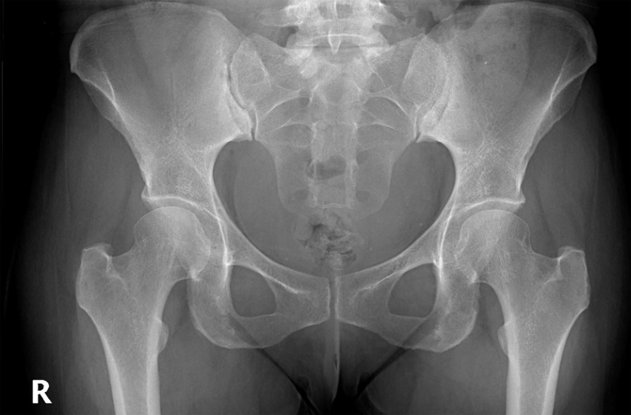
A 45-year-old woman initially presented to our orthopaedic clinic 5 weeks after an injury to the right hamstring. She was managed conservatively by a provider at an outside institution without satisfactory progress. Further workup at our institution included a pelvic radiograph, which showed no fracture or dislocations and only mild degenerative changes within the hip joint (Fig. 1). Her magnetic resonance imaging (MRI) scan revealed a 3-tendon tear of the right proximal hamstring with 3 cm of retraction. Given that she was an active individual and underwent failed conservative measures such as physical therapy and activity modification, the patient was deemed appropriate for surgical intervention.
One week later, the patient underwent open right proximal hamstring 3-anchor repair and sciatic nerve neurolysis. The procedure was uncomplicated. She reported good progress at her 3-week, 6-week, and 3-month postoperative appointments. The incision healed nicely without signs of infection. The patient was compliant with physical therapy, and, by 3 months, she had regained 4/5 hamstring strength and was able to actively flex the knee to 110°.
Six months after the operation, the patient returned to our clinic reporting worsening pain over the surgical site after slipping 5 days earlier. She described bruising and swelling in the region of the previous injury, and she was concerned that she had reinjured the hamstring. She reported that, before this episode, she had been doing extremely well and was back to nearly all activities. On examination, the incisions were well-healed and no ecchymosis was observed. There was no palpable defect along the proximal hamstring tendon or at the right ischial tuberosity, and the patient maintained her baseline 4+/5 hamstring strength on the injured side. No masses or other abnormalities were observed on examination. The patient also first mentioned at this visit that the contralateral hip had been bothering her for approximately 2 years. History and physical examination were consistent with external snapping hip.
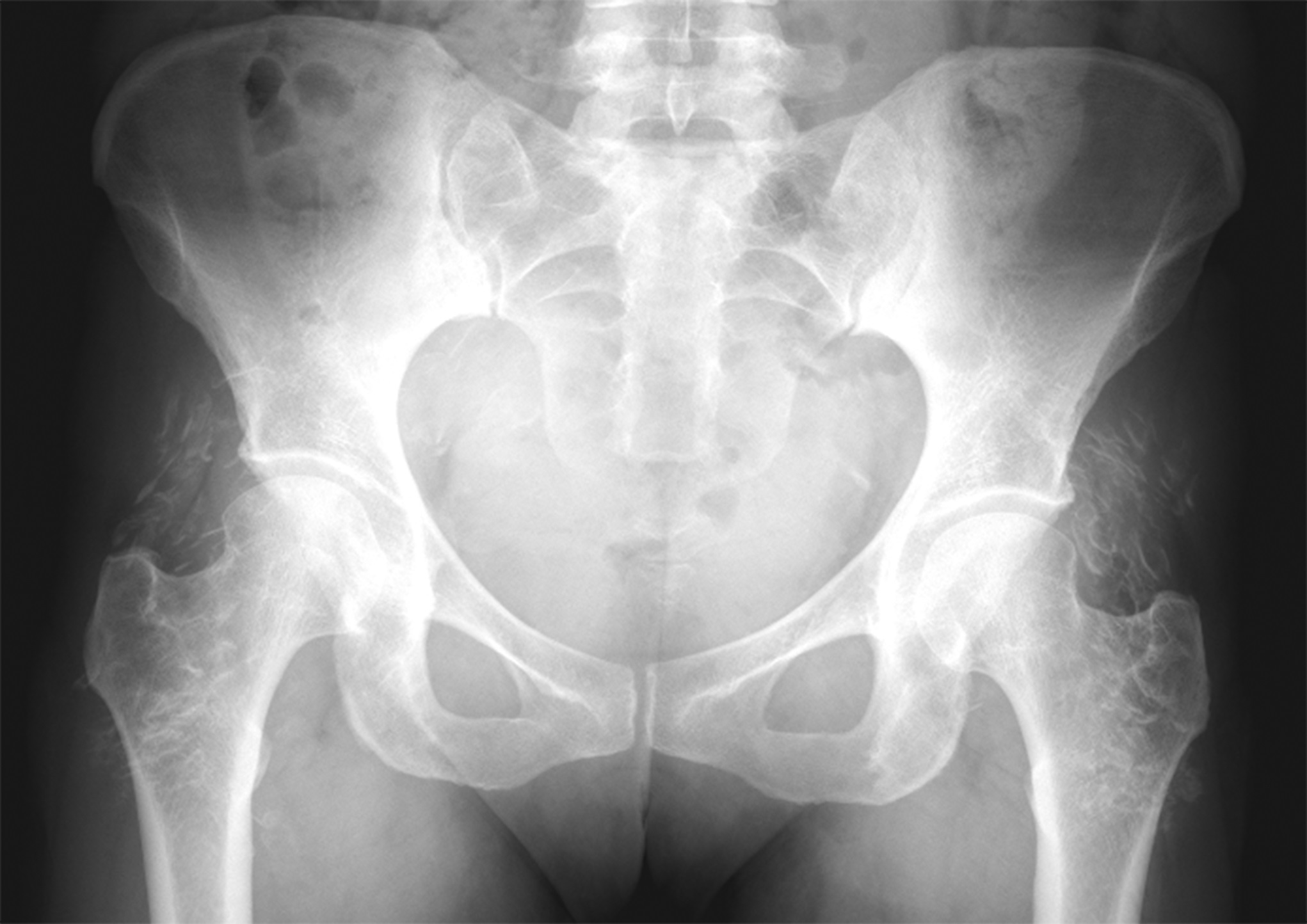
Anteroposterior pelvic radiograph revealed symmetric densities within the soft tissue around the bilateral hips. This was initially suspected to be a radiographic artifact. Repeat radiographs were made, which showed the same symmetric opacities in the abductor region of both hips (Fig. 2). Additional history gathered from the patient revealed no plausible explanation for the radiographic findings, and examination of the area was unremarkable. The patient and provider’s concern and curiosity with regard to the unique appearance on imaging prompted further investigation, and, thus, colleagues in orthopaedic oncology and radiology were consulted. Unfortunately, no definitive explanation for the radiographic findings could be provided. Extensive laboratory work was ordered to investigate possible systemic causes of calcium deposition within the soft tissues including complete blood count with differential, comprehensive metabolic panel, serum protein electrophoresis, thyroid stimulating hormone, uric acid, 25-hydroxy vitamin D, calcium, erythrocyte sedimentation rate, C-reactive protein, and rheumatoid factor. With regard to the hamstring injury, examination suggested no evidence of a retear, so the patient was given a prescription for physical therapy to help to improve her flexibility and strength. Follow-up appointments for both the radiological findings and the hamstring injury were scheduled.
The day after her appointment, the patient called to inform us that she had received bilateral gluteal injections with Radiesse (hydroxyapatite; Merz Aesthetics) for cosmetic purposes 3 months earlier. The radiographic changes were then interpreted as residual hydroxyapatite from her cosmetic injections. After recognizing that the deposits seen on the previous radiograph represented injected hydroxyapatite, additional laboratory tests and MRI scans were cancelled.
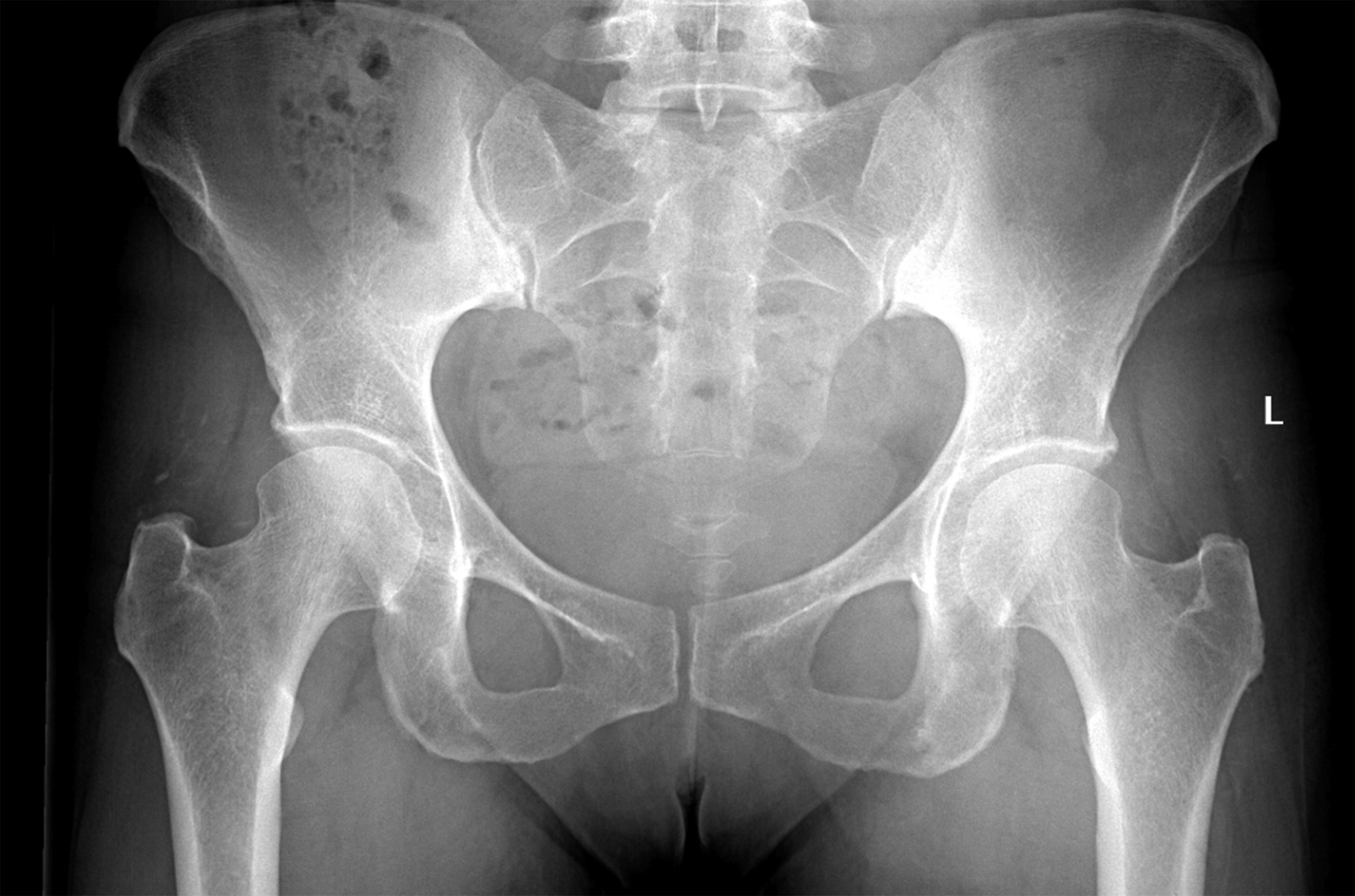
Eight months after the visit resulting in the anomalous radiograph, the patient returned to the clinic for follow-up. Pelvic radiographs showed near-complete resolution of the densities from her cosmetic injections, with some residual deposits present in the right hip abductor area (Fig. 3).
Examination of the right lower extremity revealed no palpable defect at the ischial tuberosity, 5/5 strength in the hamstrings, and no tenderness to palpation at the origin site. At the follow-up 2 years after the initial surgical procedures, the patient was doing well with no further symptoms in the right hip. At the most recent follow-up, she still had pain in the left hip. An MRI scan was ordered. However, because of the COVID-19 pandemic, the patient did not complete the advanced imaging.
Proceed to Discussion >>Reference: Holden P, Kanski G, Coyner KJ. Radiographic appearance of cosmetic gluteal injections in a patient with proximal hamstring tear: a case report. JBJS Case Connect. 2021 Dec 15;11(4):e21.00095.
Radiesse gained U.S. Food & Drug Administration (FDA) approval in 2006 for the treatment of facial fat loss (lipoatrophy) in people with human immunodeficiency virus (HIV) and to correct moderate to severe facial wrinkles and folds. The injection is meant to add volume to the facial tissue to restore a smoother appearance to the skin in the short term. It works in the long term by enhancing the production of collagen around the injection site. In 2015, it was also approved for the correction of volume loss in the dorsum of the hands, but, at the time of this writing, it was not FDA-approved for gluteal injections.
Calcium hydroxylapatite is essentially the same mineral component found in bone and teeth and can therefore be visualized on radiological imaging. An earlier version of Radiesse called Radiance FN has been FDA-approved for use as a radiological contrast agent since 2001. The original FDA filing for Radiesse included a precaution that addressed the radiopaque property of the material and urged that all patients be informed of this property so that they can disclose the information to their providers and radiologists. The filing also claimed that there was no indication that Radiesse masked abnormal tissues or was identified as tumors in computed tomography (CT) scans, although subsequent literature has indicated that it is a potential cause of false-positive imaging studies.
The material’s appearance on facial radiographs has previously been described as bilateral, amorphous clusters of discrete radiopacities with a density comparable with cancellous bone. It is hyperattenuating on CT and generally follows a distribution within fascial planes, distinct from surrounding bone. These descriptions are consistent with the findings outlined in our case, although it is important to note that the radiographic appearance may vary with time as the material is resorbed. To the best of our knowledge, Radiesse is the only dermal filler currently in use that demonstrates this radiopaque quality. However, it is important to keep in mind that calcification reactions to other, potentially unknown types of dermal fillers may also present as amorphous opacities on imaging.
Autologous fat grafting for gluteal augmentation has also prompted referrals to orthopaedic oncology similar to this case. Lugo-Pico et al. described 3 such cases in patients with a history of autologous fat grafting for gluteal augmentation spanning from 3 months to 6 years before presentation. They found that these patients presented with painless or painful masses in the lower extremity and gluteal areas and described characteristic MRI findings including heterogeneous masses with floating fat globules and a band-like pattern of fat deposition within the muscle resembling tiger stripes.
One must also consider that patients may receive dermal injections for cosmetic purposes from unlicensed professionals with non-FDA-approved injectables. For example, radiographic reports of scattered, nodular soft-tissue masses representing silicone granulomas have been described in patients who received nonmedical-grade liquid silicone injections. A more thorough discussion of the radiographic appearance of various cosmetic injections and implants has been described elsewhere.
With the increasing popularity of cosmetic injections, it is important to be able to distinguish the radiological appearance of cosmetic injection materials from other, potentially serious, pathologic findings. In addition, it is important to keep in mind that, as seen in our case, the radiodensity of the filler may change over time as the material is resorbed. The presence of bilateral, ill-defined radiopaque masses in soft tissue, without a clear etiology, should prompt the clinician to inquire about cosmetic injections and, if the patient did receive these injections, investigate the type of injection used.
Reference: Holden P, Kanski G, Coyner KJ. Radiographic appearance of cosmetic gluteal injections in a patient with proximal hamstring tear: a case report. JBJS Case Connect. 2021 Dec 15;11(4):e21.00095.
What is the diagnosis?
Tumoral calcinosis
Heterotopic ossification
Myositis ossificans
Hydroxyapatite gluteal injections
Cavernous hemangiomas with calcification