 Fig. 1-A
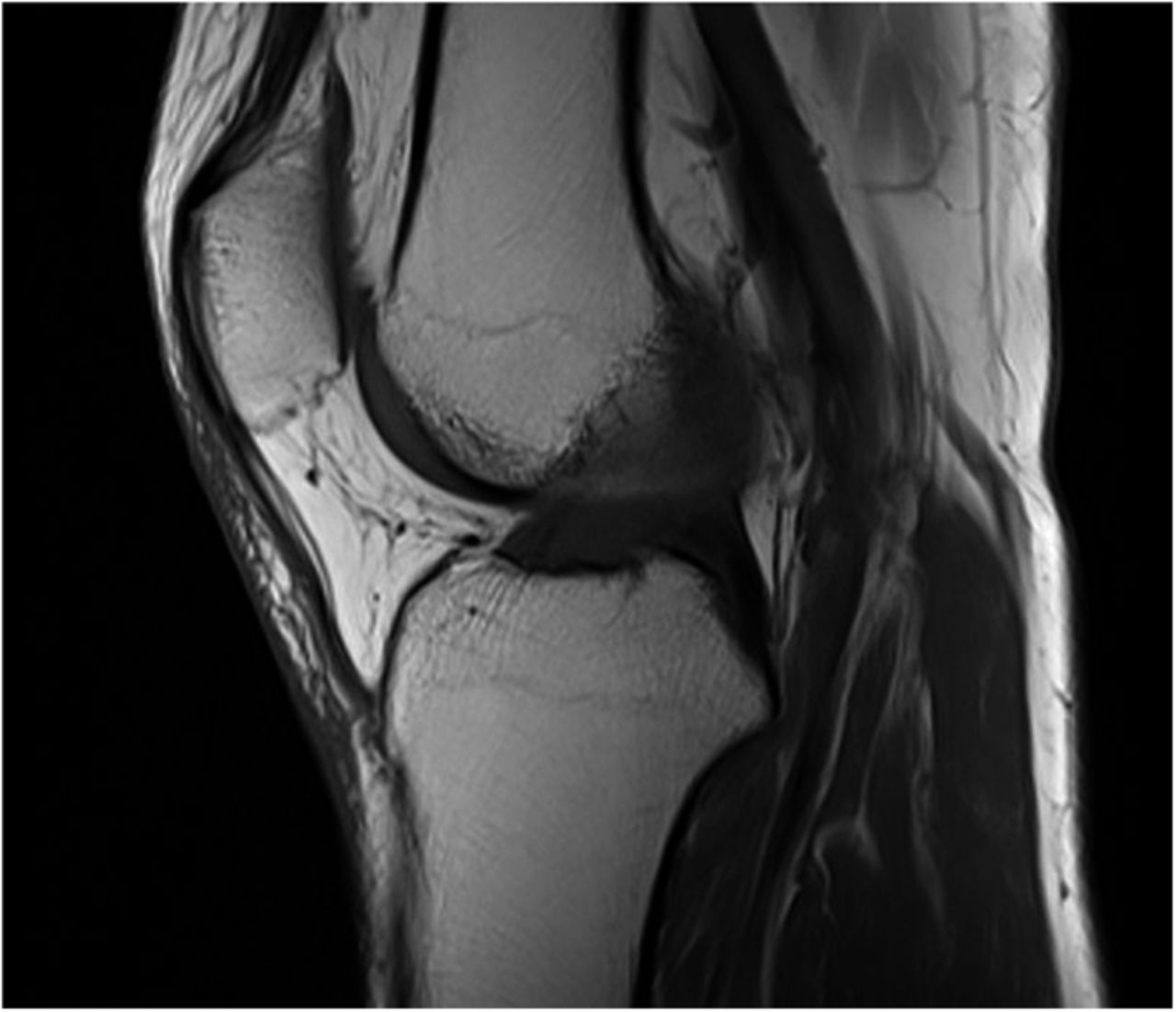
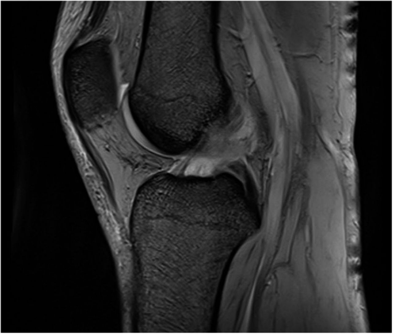
 Fig. 1-B
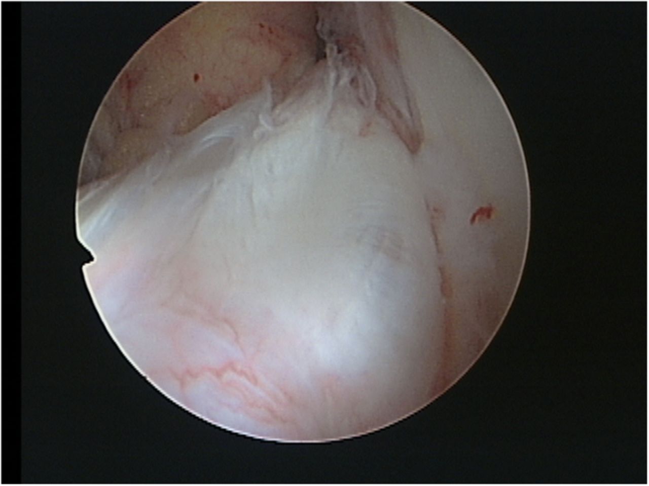
 Fig. 2-A
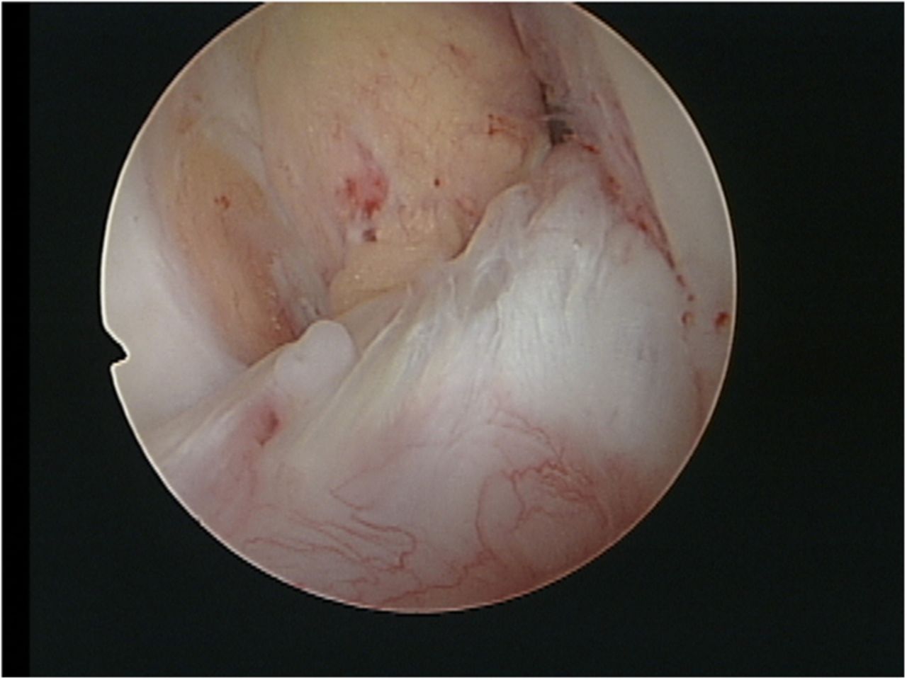
 Fig. 2-B
 Fig. 2-C
 Fig. 3
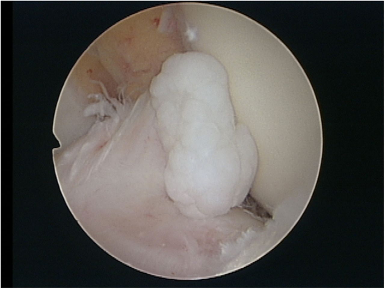
A forty-seven-year-old man with no history of substantial trauma presented with progressive pain in the left knee and limited range of motion. He walked with a limp and had difficulty squatting and climbing up and down stairs. On examination, the knee was found to have mild effusion. Extension of the knee was limited to −15°, with terminal pain. The McMurray test was negative. Ligamentous injuries were not detected; the Lachman test, pivot-shift test, anterior and posterior drawer tests, and varus and valgus stress tests were all negative. The numeric rating scale score for assessment of pain intensity was 8 (range, 0 [best] to 10 [worst]), and the Lysholm knee scale score was 55 points (range, 0 [worst] to 100 [best]). Radiographs showed no abnormalities. Magnetic resonance imaging (MRI) revealed swelling of the anterior cruciate ligament (ACL) and an intraligamentous mass with low signal intensity on T1-weighted imaging and high signal intensity on T2-weighted imaging (Figs. 1-A and 1-B). The symptoms had persisted for more than three months and had not responded to physical therapy; an arthroscopic examination was performed. We found moderate proliferation of the synovial tissue in the joint, which was removed with use of a shaver. There were no free bodies in the joint cavity. However, there was obvious thickening of the ACL, which impinged on the femoral intercondylar notch (Fig. 2-A). No mass was found outside the ACL.
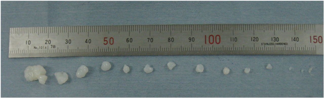
Aspiration of the lesion in the ACL was ineffective. A longitudinal incision was then made in the anterior part of the ACL, and approximately one dozen chondral bodies were expelled from the ACL (Figs. 2-B, 2-C, and 3). After the chondral bodies were removed, the ACL became decompressed, and we ascertained that there was no notch impingement. The posterior cruciate ligament (PCL) was intact, and there was no meniscal or chondral lesion. The histological diagnosis was synovial chondromatosis. The patient regained full range of motion of the left knee and was pain-free immediately after the operation. The Lachman test, pivot-shift test, and anterior and posterior drawer tests were all negative. At the eighteen-month follow-up, the patient had maintained full range of motion of the knee, without pain or instability. The numeric rating scale score had decreased to 1, and the Lysholm knee scale score had improved to 95 points.
Proceed to Discussion >>Reference: Mio K, Tamura K, Nemoto K. Intraligamentous synovial chondromatosis of the anterior cruciate ligament: a case report. JBJS Case Connector. 2014 Jan 08;4(1):e4.
To our knowledge, only three cases of synovial chondromatosis of the ACL have been reported, along with one case of synovial chondromatosis in the PCL and one case of synovial chondromatosis in both ligaments. In those cases, the chondral bodies were situated around the cruciate ligaments. To our knowledge, the case described here represents the first reported case of synovial chondromatosis located in the intraligamentous area of the ACL. Synovial chondromatosis is defined as chondroid metaplasia of the subintimal tissue. Synovial chondromatosis usually involves the entire joint. In our patient, however, the lesion was restricted to the intraligamentous area of the ACL. This is an extremely rare condition, and the diagnosis was nearly impossible before the surgical procedure. Because the lesion was in the ACL, the symptoms (pain and loss of range of motion) were nonspecific. There was no “catching” of the knee, which is often caused by free bodies resulting from synovial chondromatosis. Radiographs revealed no abnormalities because the chondral bodies were not calcified. MRI was of some help because it suggested the presence of a mass in the ACL. However, the pieces of the chondral bodies were not visible on MRI scans, likely because they had a signal intensity similar to that of joint fluid. The MRI findings in the present case were similar to those associated with mucoid degeneration of the ACL, which is marked by ACL thickening and the presence of a ganglion cyst. The ACL thickening in the case of our patient was due to the presence of chondral bodies in the intraligamentous area of the ACL, unlike previous cases of mucoid degeneration, in which ACL thickening was caused by a mucinous degenerative change of the connective tissue. Because the chondral bodies were not visible on MRI scans, we could not distinguish the present case from the mucoid degeneration of the ACL at this point. An unusual feature of the present case was that a definitive diagnosis was impossible preoperatively. The use of arthroscopy was indispensable. We made a longitudinal incision in the anterior part of the ACL, where we found approximately one dozen chondral bodies, which we extracted. We were able to preserve the ACL, resulting in excellent function of the knee without pain or instability. In contrast, Chung et al. reported a case in which ACL resection during the removal of osteochondral bodies caused knee instability. In another case, the open approach was required after an arthroscopic operation in order to deal with the posterior recess. In the case of our patient, the lesion was located in the ACL, and an arthroscopic operation was effective. Because the patient recovered immediately after removal of the chondral bodies, the mass in the ACL was presumed to be responsible for the pain and loss of range of motion. The chondral bodies that were localized in the intraligamentous area of the ACL had apparently caused the swelling of the ACL, which then had impinged on the femoral intercondylar notch, producing the symptoms.
Reference: Mio K, Tamura K, Nemoto K. Intraligamentous synovial chondromatosis of the anterior cruciate ligament: a case report. JBJS Case Connector. 2014 Jan 08;4(1):e4.
Posterior horn of the lateral meniscus tear that is unstable
Pigmented villonodular synovitis
Unstable osteochondritis dissecans lesion of the distal part of the femur
Intra-articular chondrosarcoma
Synovial chondromatosis of the knee