An Abnormal Radiograph After Volar Locking Plate Fixation of a Distal Radial Fracture
August 4, 2010
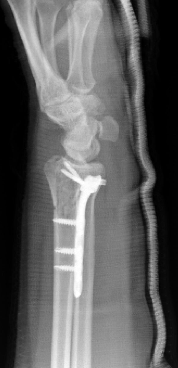
 Fig. 2
 Fig. 2
 Fig. 1
A forty-eight-year-old woman fell on ice and sustained a closed extra-articular distal radial fracture with 40° of dorsal angulation and dorsal comminution. She underwent open reduction and internal fixation with a Distal Volar Radius (DVR) locking plate (Hand Innovations, Miami, Florida). The proximal row of the DVR plate was filled with locking screws and the distal row was not used. Intraoperative fluoroscopy was employed but was discontinued after placement of the screws. Postoperative radiographs showed this configuration of the fixation device.
[review images below, then proceed to the Discussion tab]
Proceed to Discussion >>Reference: Bhattacharyya T, Wadgaonkar AD. Inadvertent retention of angled drill guides after volar locking plate fixation of distal radial fractures. A report of three cases. J Bone Joint Surg Am. 2008;90:401-3.
Volar locking plates have revolutionized the care of distal radial fractures. Our case is one of three that we have seen that represent a potential pitfall associated with one type of locking plate, in addition to the pitfalls already reported. Preloading the plate with angled drill guides allows for quick insertion of the locking screws. However, if not all of the potential holes are filled with a screw, care must be taken to remove the drill guides before closure. Because this complication occurred on three separate occasions and affected the patients of three different surgeons, it is unlikely to be due to simple error and represents a small risk that is associated with such newer implant designs. These three surgeons collectively repaired 131 distal radial fractures with this plate design during the same time period. Thus, while the rate of retention (2.3%) is low, additional routine checks must be performed to prevent the complication of retained angled drill guides. In the first two of our three patients, the event may have been avoided by making postoperative radiographs prior to leaving the operating room. However, this practice has become rarer as operating-room time has become tighter and cost-control pressures on surgeons have increased. The ability to record fluoroscopy films in the digital film library has further decreased the need for making traditional radiographs in the operating room. Retained drill guides had not been noted on any of the official radiology reports for the three patients. Another possible method to avoid retained angled drill guides is to fill all of the screw holes in the volar locking plate. Filling all screw holes necessitates removal of the drill guides prior to measuring and screw insertion. However, it is often unnecessary to provide additional fixation, especially in the distal row of the plate where the risk of intra-articular penetration increases with additional screws. Use of a separate angled drill guide that screws into the plate (rather than being preloaded) would prevent this complication but would increase operating-room time. On the basis of these three cases, we recommend that when a locking-plate drill guide is inadvertently left in the patient, the guide should be removed as soon as possible so as to prevent flexor tendon rupture. Although flexor tendon rupture occurred in only one of our three patients, it is a clear risk and the potential for morbidity is considerable. We also strongly recommend an additional step of palpating the plate for retained drill guides prior to closure, and/or obtaining traditional postoperative radiographs in the operating room. With these measures in place, we believe that inadvertent retention of drill guides can be entirely prevented.
Reference: Bhattacharyya T, Wadgaonkar AD. Inadvertent retention of angled drill guides after volar locking plate fixation of distal radial fractures. A report of three cases. J Bone Joint Surg Am. 2008;90:401-3.

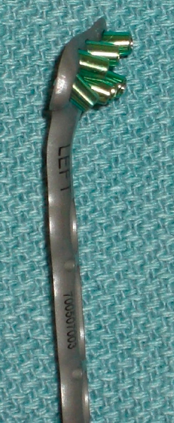
Retained angled drill guides
Broken distal screws
Normal plate and screw positions