An Unusual Cause of Late Pain and Effusion After Total Knee Arthroplasty
August 18, 2010

 Fig. 1
 Fig. 1
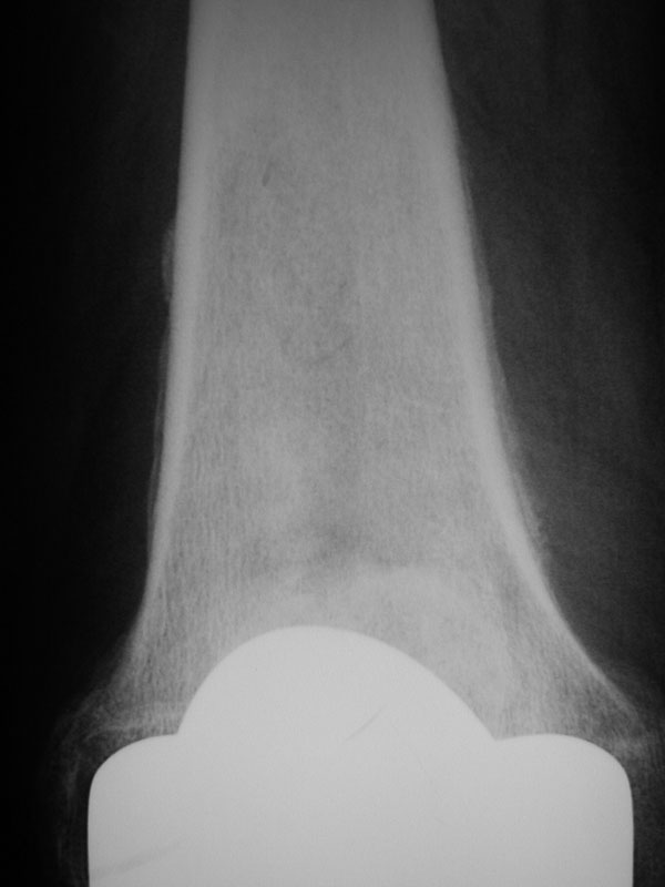
A seventy-seven-year-old woman presented in early February 2007 with a two-month history of pain and swelling in both knees. She had undergone a bilateral staged total knee arthroplasty with cement for the treatment of osteoarthritis in January and November 2001, without complications. Routine follow-up examinations and radiographs in December 2005 showed unremarkable findings. The onset of the pain and swelling was insidious, and there was no antecedent trauma. The patient was able to walk outdoors with a cane and did not report any fever or chills. Nine months before the examination for the pain and swelling in the knees, hoarseness had developed and a chest radiograph made at another institution showed a left hilar mass. Mediastinoscopy and lymph node biopsy at that institution showed small-cell carcinoma. The patient had a vocal cord injection for the hoarseness, and a workup for metastases revealed negative findings. After extensive discussion of treatment options with her oncologist, the patient decided against radiation or chemotherapy and wanted only supportive care. On examination for the pain and swelling in the knees, the patient was afebrile and walked without a limp. The knees were both warm, without erythema, and both had effusions. There was tenderness to deep palpation about the distal parts of both femora. The range of motion was from full extension to 120° of flexion bilaterally. There was no instability or crepitus. Inspection of the hands showed clubbing of multiple digits. There were no radiolucent lines and no evidence of osteolysis. The hemoglobin level was 122 g/L (normal, 120 to 160 g/L), the white blood-cell count was 12.3 × 10⁹/L (normal, 4.5 to 1.0 × 10⁹/L), the erythrocyte sedimentation rate was 82 mm/hr (normal, 0 to 30 mm/hr), and the C-reactive protein level was 6.3 mg/dL (normal, 0 to 100 mg/dL). As infection was suspected, the sites of the total knee arthroplasties were sequentially aspirated. Each knee had 20 mL of clear yellow fluid with a total nucleated cell count of 124/mm3 and 1% neutrophils, 76% lymphocytes, and 23% other cells. No crystals were seen. There was no growth after seven days of aerobic or anaerobic culture of the aspirate from either knee. An anteroposterior radiograph of the knee is presented in Figure 1.
Radiographs showed periosteal new bone formation at the medial and lateral parts of the distal femoral metaphysis bilaterally (Fig. 1). There were no radiolucent lines and no evidence of osteolysis. With the exclusion of infection, loosening, instability, and crystalline synovitis, we made the presumptive diagnosis of pulmonary hypertrophic osteoarthropathy of the distal parts of both femora. Indomethacin (25 mg three times per day) was prescribed, and there was complete relief of the pain and effusions within one week. The patient was evaluated six weeks later and, with the continued absence of symptoms, it was decided that the indomethacin therapy would be continued indefinitely.
Proceed to Discussion >>Reference: Meeker J, Lachiewicz PF. An unusual cause of late pain and effusion after total knee arthroplasty: pulmonary hypertrophic osteoarthropathy. A case report. J Bone Joint Surg Am. 2008;90:390-2.
To our knowledge, this is the first report of pulmonary hypertrophic osteoarthropathy occurring in a patient who had had a total knee arthroplasty. This disorder exists in both primary and secondary forms. Primary pulmonary hypertrophic osteoarthropathy presents in young adults with digital clubbing, periosteal reaction of the long bones, and hypertrophic skin changes. The secondary form is much more common and is associated with malignant tumors, chronic pulmonary disease, cardiac disorders, and certain gastrointestinal diseases. A review of the literature identified four reports of pulmonary hypertrophic osteoarthropathy associated with small-cell carcinoma of the lung. The symptoms of pulmonary hypertrophic osteoarthropathy can include debilitating joint pain, effusions, and clubbing of the fingers. The exact etiology of secondary pulmonary osteoarthropathy remains uncertain, but the underlying disorders probably elicit a common physiologic response. One hypothesis is that pulmonary dysfunction allows vasogenic cell fragments to escape the pulmonary circulation as a result of shunting. These particles lodge distally and evoke a metabolic response from local tissues. Vascular endothelial growth factor has been noted to be elevated in some patients with this disorder. The diagnosis of pulmonary hypertrophic osteoarthropathy requires a carefully recorded history, physical examination, and radiographs. The findings should include joint pain, effusions, and clubbing of the digits. The joint fluid of patients with this disorder has fewer than 500 nucleated cells per high-powered field despite an elevated erythrocyte sedimentation rate. The characteristic radiographic finding is a periosteal reaction that increases in intensity during the duration of the disease process. The tibia and fibula are the most common locations in the extra-axial skeleton, and the distal part of the femur is the second most common site of involvement. The bone scan is the most sensitive test for identifying this disorder, and it can detect subclinical cases. A recent study demonstrated the utility of a positron emission tomography/computerized tomography scan for the diagnosis of pulmonary osteoarthropathy. The mainstay of treatment of secondary pulmonary hypertrophic osteoarthropathy involves the management of the underlying disorder. There have been several reports of a decrease in symptoms following resection, chemotherapy, or irradiation of an underlying malignant tumor. If it is not feasible to treat the underlying disorder, then relief of bone pain and joint effusion can be obtained with indomethacin or other nonsteroidal anti-inflammatory medication. There have been reports of effective treatment with pamidronate and octreotide. Our patient's history of small-cell carcinoma of the lung, the physical findings of clubbing and joint effusion, and the radiographic changes in the distal parts of the femora suggested the diagnosis of pulmonary hypertrophic osteoarthropathy. The diagnosis in this patient helped to avoid unnecessary surgery and led to effective treatment of symptoms with indomethacin rather than strong analgesics.
Reference: Meeker J, Lachiewicz PF. An unusual cause of late pain and effusion after total knee arthroplasty: pulmonary hypertrophic osteoarthropathy. A case report. J Bone Joint Surg Am. 2008;90:390-2.
Infection of total knee arthroplasty site and distal part of the femur
Osteolysis from wear debris after total knee arthroplasty
Pulmonary hypertrophic osteoarthropathy
Metastatic disease of the distal part of the femur